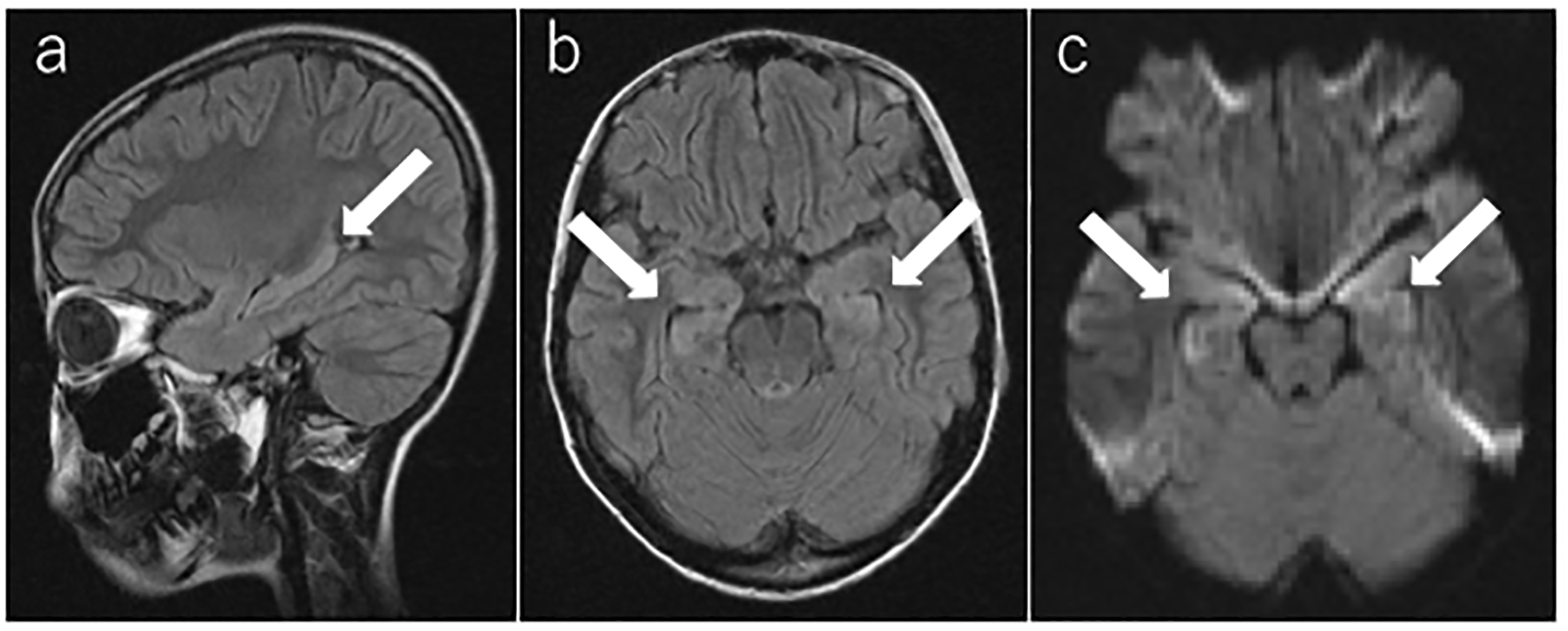
Figure 1. Brain magnetic resonance imaging. (a, b) T2-weighted fluid-attenuated inversion recovery image and (c) diffusion-weighted image on the second day of admission (day 14). The arrow shows the high-intensity areas in the bilateral medial temporal lobes and hippocampus.
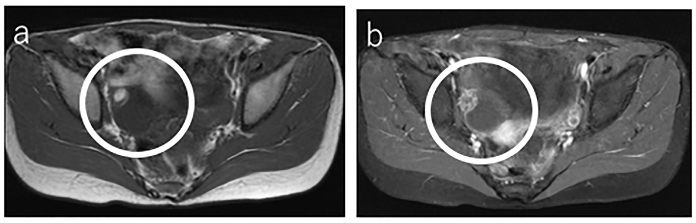
Figure 2. Abdominal magnetic resonance imaging. (a) T1-weighted fluid-attenuated inversion recovery image on day 31 showed the right ovarian tumor with a high-intensity cystic lesion. (b) Fat-saturated gadolinium-enhanced T1-weighted fluid-attenuated inversion recovery image showed a low-intensity lesion.
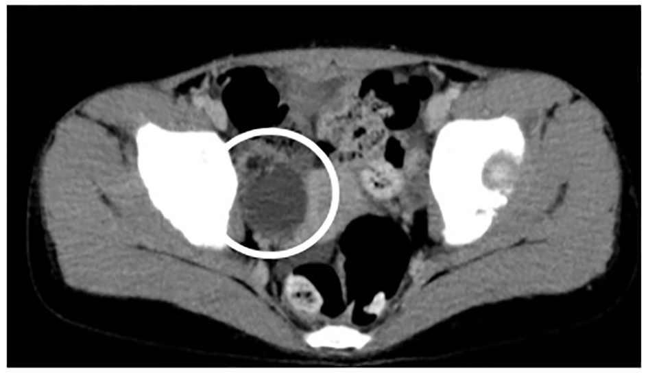
Figure 3. Enhanced abdominal computed tomography. On day 35, computed tomography showed the right ovarian tumor, including calcific and fatty components.
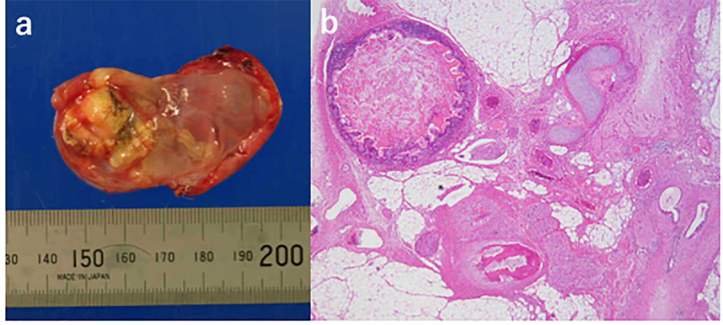
Figure 4. Pathological examination of the resected right ovarian tumor. (a) Macroscopic image of the right ovarian tumor after incision to reveal some hair (mature ovarian teratoma). A centimeter scale is shown under the specimen. (b) Histopathological image of the right ovarian tumor shows heterologous elements including central nervous system tissue, cerebellar tissue, choroid plexus, ganglion, skin, fat, sebaceous glands, hair follicles, cartilage, bone, and intestinal epithelia.