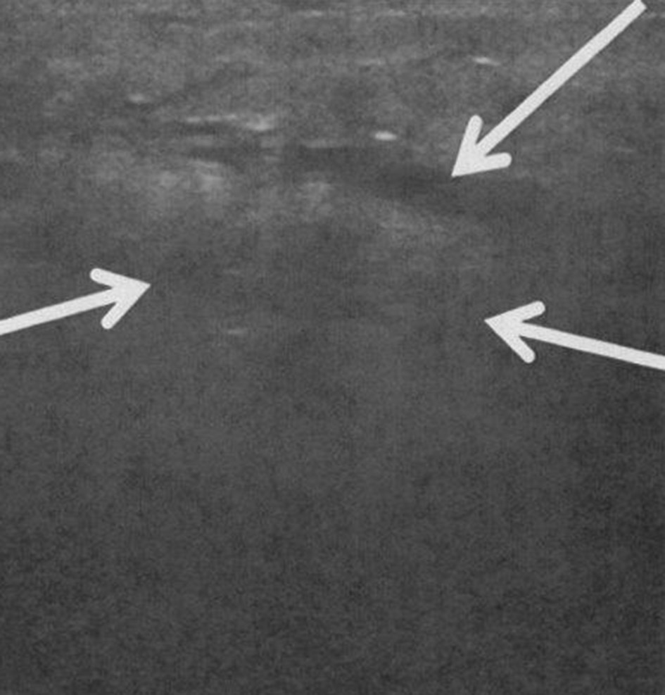
Figure 1. Ultrasound (US) scan of right parotid gland. Longitudinal US image of the right parotid region showing ill-defined tissue with surrounding edema (arrow). No echogenic stones, gas or dilated parotid ducts are seen.
| International Journal of Clinical Pediatrics, ISSN 1927-1255 print, 1927-1263 online, Open Access |
| Article copyright, the authors; Journal compilation copyright, Int J Clin Pediatr and Elmer Press Inc |
| Journal website http://www.theijcp.org |
Case Report
Volume 7, Number 3, September 2018, pages 36-38
Non-Infectious Parotitis in Infants with Severe Bronchopulmonary Dysplasia: A Case Series
Figures
Table
| Patient | 1 | 2 | 3 | 4 |
|---|---|---|---|---|
| *PMA: post-menstrual age; **temperature ≥ 38°C; ***white blood cell count > 11,000; ****US: ultrasound; *****CT: computed tomography scan; ******MRI: magnetic resonance imaging. | ||||
| Gender | Female | Female | Male | Female |
| Gestational age (weeks) | 24 | 25 | 24 | 29 |
| Birth weight (g) | 600 | 640 | 830 | 1,005 |
| Maternal age (years) | 22 | 25 | 30 | 29 |
| Mode of delivery (cesarean section (CS)) | CS | CS | CS | CS |
| Apgar scores (1 and 5 min) | 6, 6 | 5, 6 | 1, 3 | 6, 8 |
| Age at tracheostomy (weeks, PMA*) | 41 | 43 | 36 | 48 |
| Age at diagnosis of parotitis (weeks, PMA*) | 42 | 50 | 44 | 52 |
| Location (unilateral/bilateral) | Bilateral | Bilateral | Unilateral | Bilateral |
| Fever at time of parotitis** (yes/no) | No | No | Yes | Yes |
| Leukocytosis at time of parotitis*** (yes/no) | No | No | Yes | Yes |
| Number of ventilator days prior to parotitis | 125 | 175 | 135 | 160 |
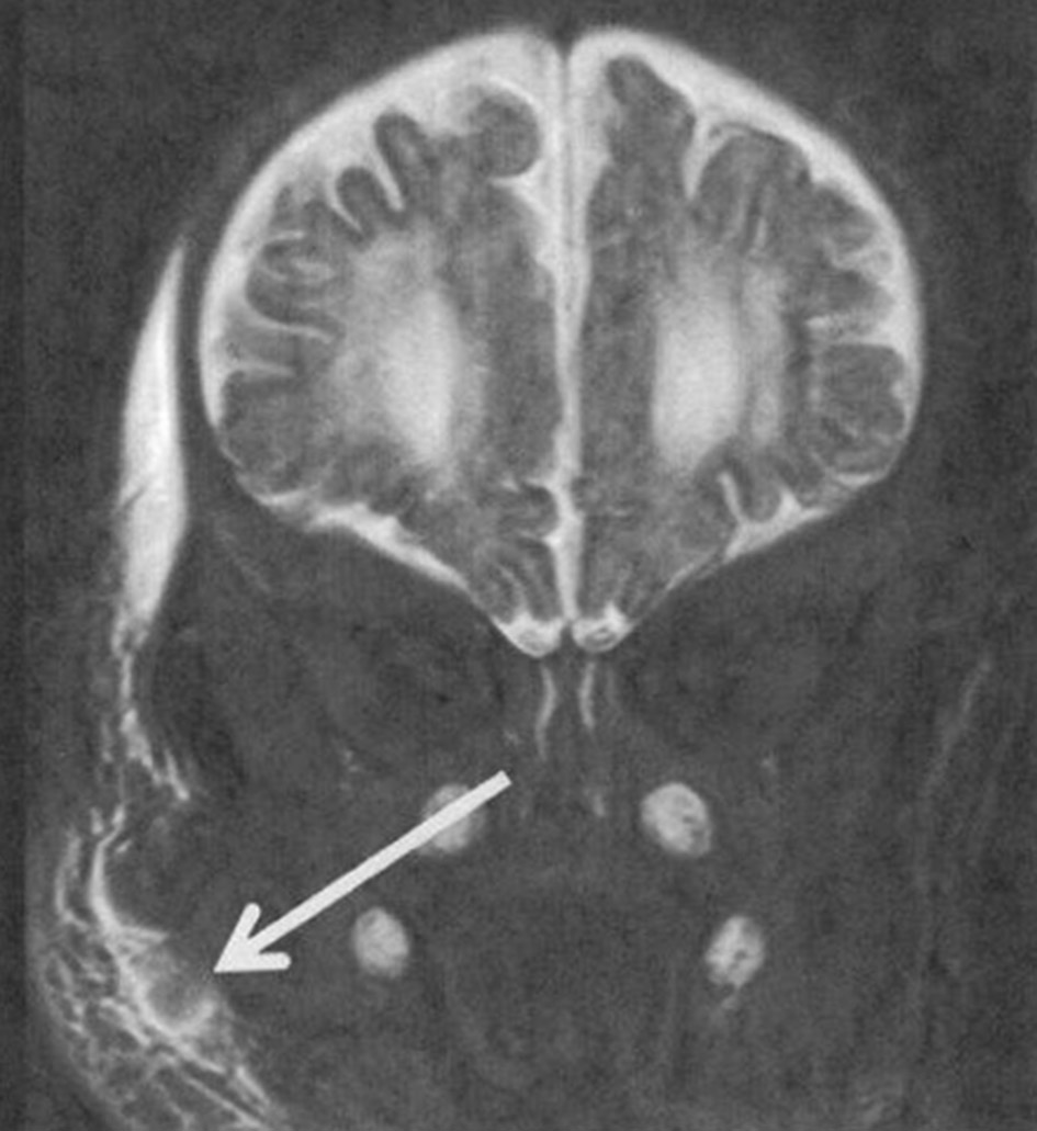
| Imaging study for parotitis (US****, CT*****, MRI******) | US/CT | MRI | CT | US |
| Antibiotics treatment for parotitis (yes/no) | Yes | No | Yes | No |
| Recurrence of parotitis (yes/no) | Yes | Yes | No | No |
| Complications from parotitis (yes/no) | No | No | No | No |