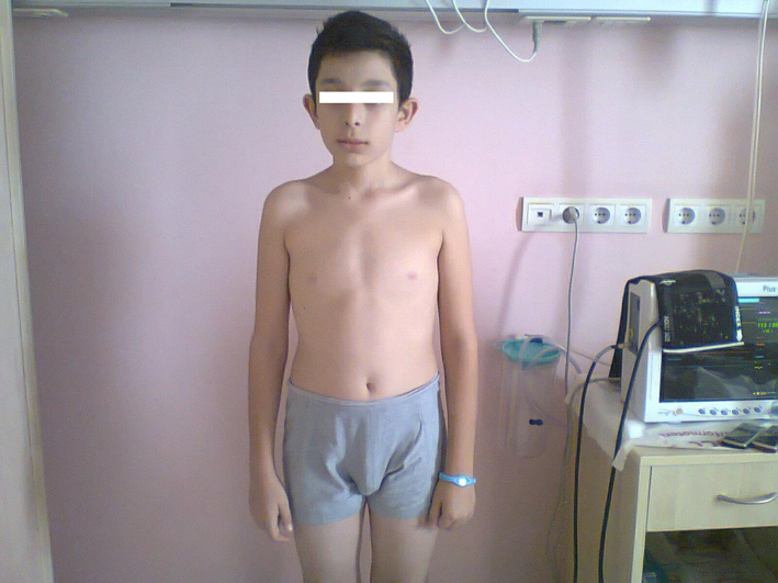
Figure 1. Features of patient.
| International Journal of Clinical Pediatrics, ISSN 1927-1255 print, 1927-1263 online, Open Access |
| Article copyright, the authors; Journal compilation copyright, Int J Clin Pediatr and Elmer Press Inc |
| Journal website http://www.theijcp.org |
Case Report
Volume 2, Number 2, December 2013, pages 78-82
Importance of Ultrasound Imaging in Pheochromocytoma in a Child: Case Report and Review of Literature
Figures
Table
| Pre-op | Post-op | |
|---|---|---|
| Blood pressure | 170/100 mmHg | 100/80 mmHg |
| Hearth rate | 140/min | 96/min |
| Fasting blood glucose | 142 mg/dL | 86 mg/dL |
| RBC | 5.2 million/mm3 | 3.7 million/mm3 |
| WBC | 13.2 × 10³/mm3 | 5.2 × 10³/mm3 |
| ANC | 3,400 | 1,700 |
| ESR | 65/30 min | 9/30 min |
| Urinary metanephrine | 1,278 µg/24 h | 246 µg/24 h (reference: 52 - 341 µg/24 h) |
| Urinary VMA | 19.3 mg/24 h | 4 mg/24 h (reference: 3 - 9 mg/24 h) |
| Urinary epinephrine | 747 µg/24 h | 6 µg/24 h (reference: 0 - 20 µg/24 h) |
| Urinary norepinephrine | 254 µg/ 24 h | 25 µg/24 h (reference: 15 - 80 µg/24 h) |