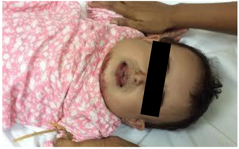
Figure 1. The patient’s picture showing facial swelling, taken about 3 - 4 h from its onset.
| International Journal of Clinical Pediatrics, ISSN 1927-1255 print, 1927-1263 online, Open Access |
| Article copyright, the authors; Journal compilation copyright, Int J Clin Pediatr and Elmer Press Inc |
| Journal website http://www.theijcp.org |
Case Report
Volume 4, Number 1, March 2015, pages 139-142
Subcutaneous Emphysema and Pneumomediastinum Following Oro-Facial First Degree Burns
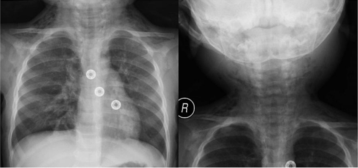
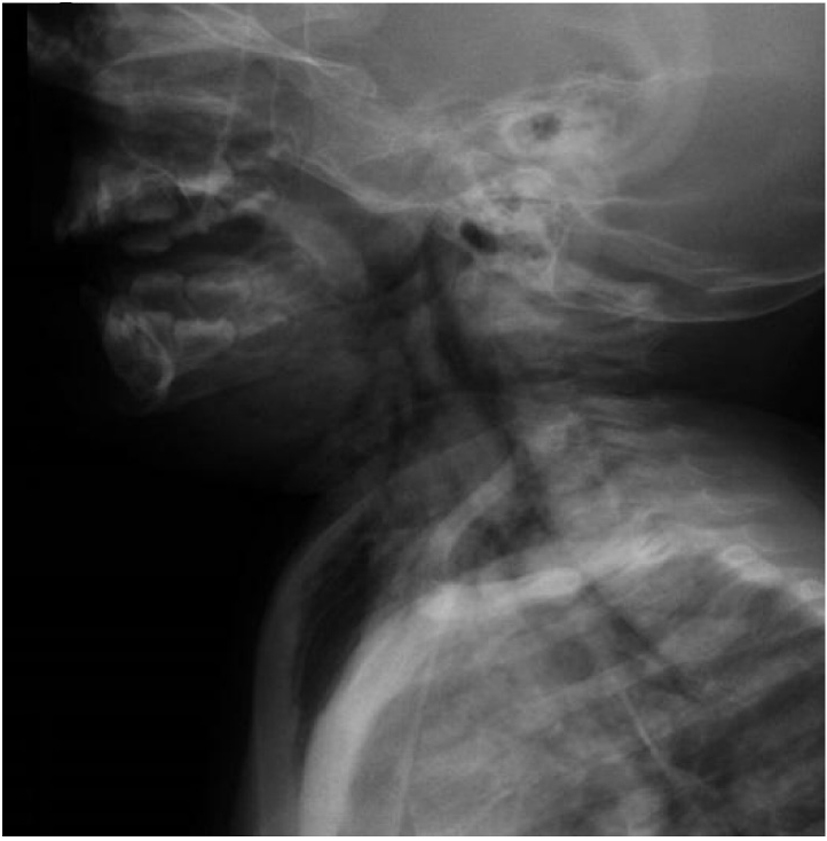
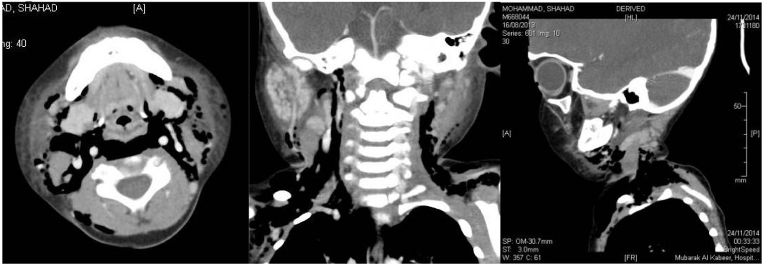
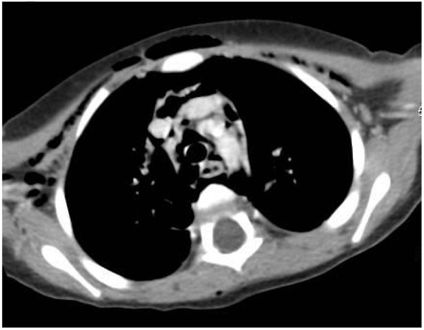
Figures