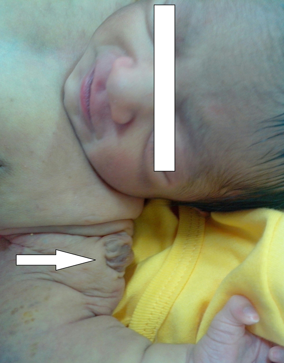
Figure 1. Photograph of the child showing papulonodular swelling over the left shoulder (white arrow).
| International Journal of Clinical Pediatrics, ISSN 1927-1255 print, 1927-1263 online, Open Access |
| Article copyright, the authors; Journal compilation copyright, Int J Clin Pediatr and Elmer Press Inc |
| Journal website http://www.theijcp.org |
Case Report
Volume 5, Number 3-4, December 2016, pages 47-50
Congenital Embryonal Rhabdomyosarcoma Presenting as a Cutaneous Nodule in a Neonate
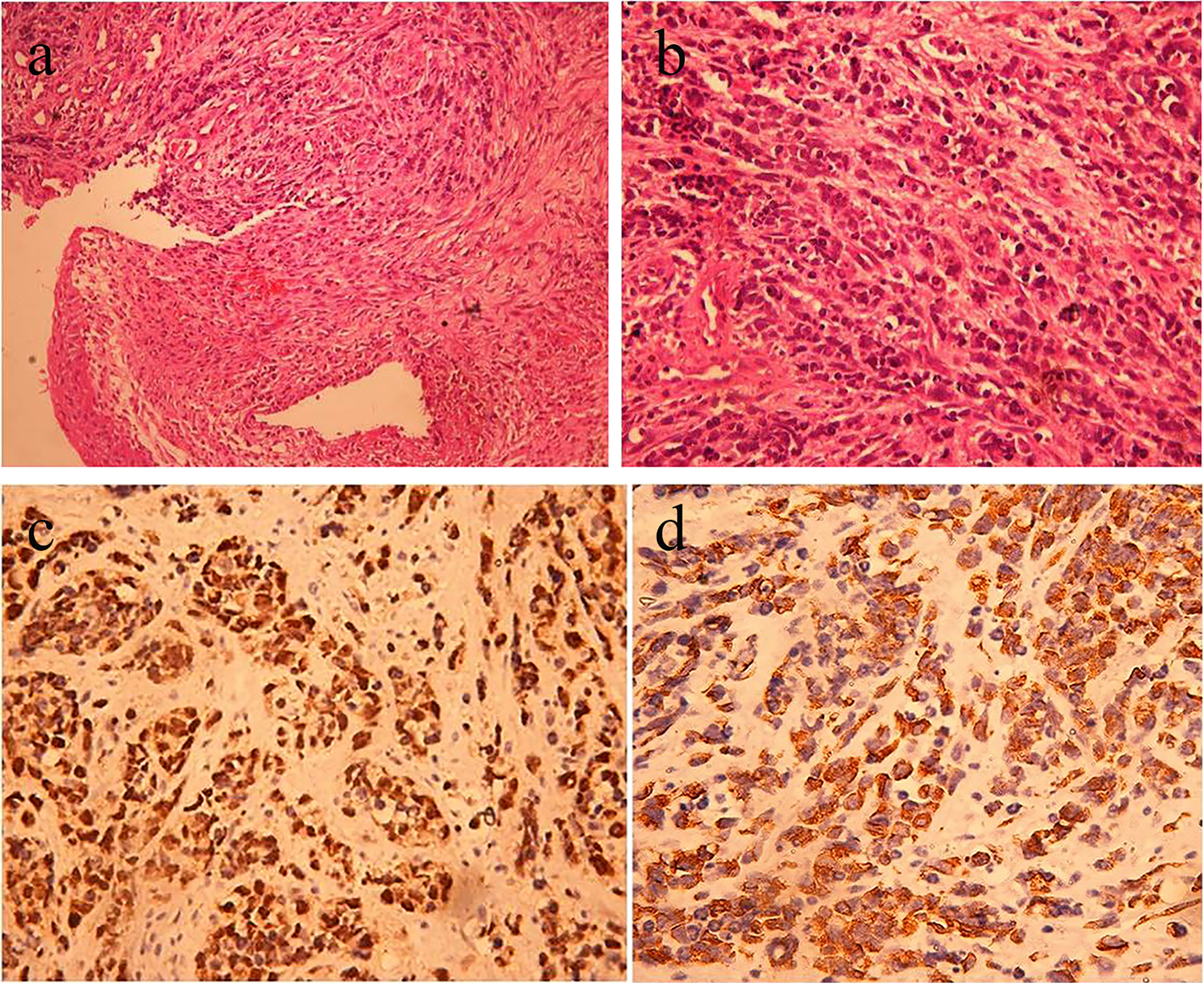
Figures