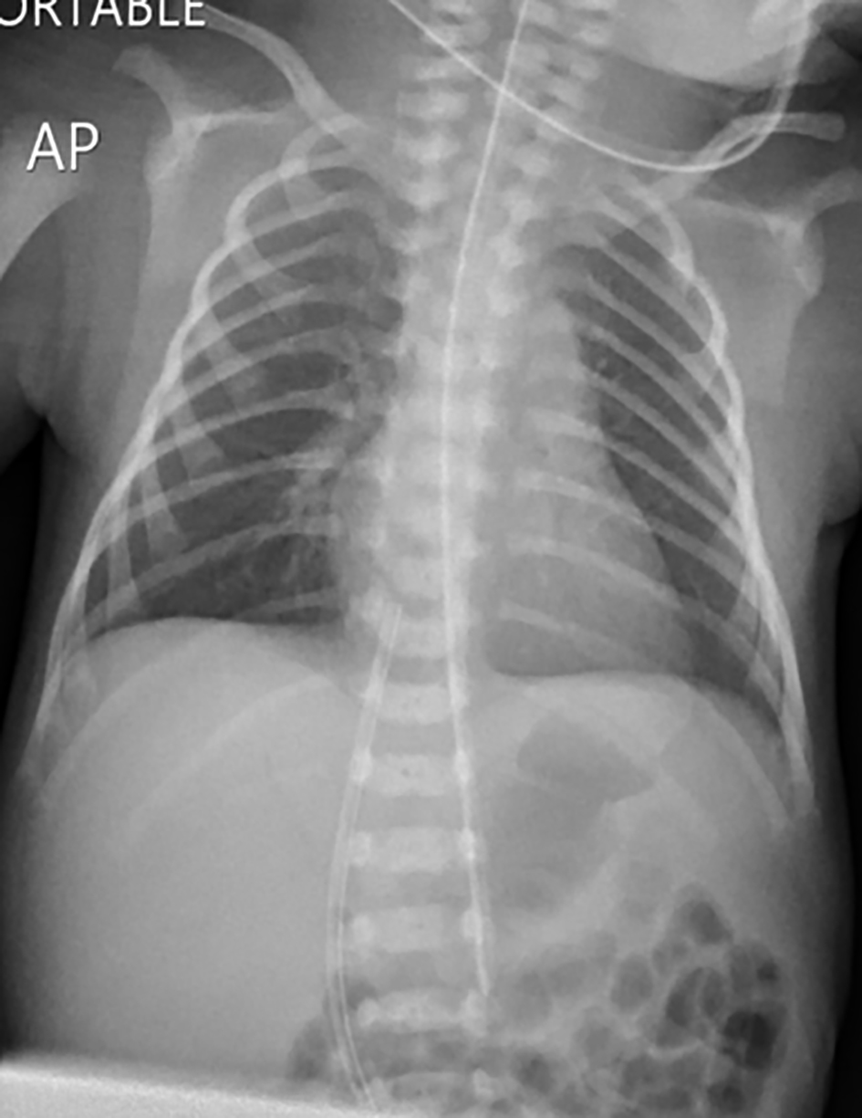
Figure 1. Normal chest and abdomen radiology on admission showing correctly placed UVC at the level of T9. UVC: umbilical venous catheter.
| International Journal of Clinical Pediatrics, ISSN 1927-1255 print, 1927-1263 online, Open Access |
| Article copyright, the authors; Journal compilation copyright, Int J Clin Pediatr and Elmer Press Inc |
| Journal website http://www.theijcp.org |
Case Report
Volume 8, Number 1, August 2019, pages 22-25
A Preterm Infant With Mild Abdominal Distension and Rising C-Reactive Protein
Figures
Table
| FBC | LFT | PT | CRP | Na | K | Cr | |||||||||
|---|---|---|---|---|---|---|---|---|---|---|---|---|---|---|---|
| WBC | Hb | Plat | TB, mg/dL | ALP, U/L | ALT, U/L | TP, g/dL | Alb, g/dL | Glob | AFP, ng/mL | ||||||
| FBC: full blood count; LFT: liver function test; WBC: white blood cells; Hb: hemoglobin; Plat: platelets; TB: total bilirubin; ALP: alkaline phosphatase; ALT: alanine transferase; TP: total protein; Alb: albumin; Glob: globulin; AFP: alpha-fetoprotein protein; PT: prothrombin time; CRP: C-reactive protein; Cr: creatinine. | |||||||||||||||
| Day 1 | 13.2 | 14.9 | 372 | 0.9 | - | - | - | - | - | - | - | 7.1 | 139 | 5 | - |
| Day 4 | 13.7 | 15.2 | 385 | 4.5 | 264 | 35 | 5.1 | 3.2 | 1.9 | - | 14 | 22 | 142 | 3.5 | 0.6 |
| Day 10 | 8.9 | 8 | 455 | 1.9 | 259 | 22 | 5.1 | 3 | 2.1 | 30,000 | 12 | 41.5 | 139 | 5.4 | 0.6 |
| Day 24 | 8 | 9.7 | 487 | 1 | 277 | 13 | 5.3 | 2.9 | 2.4 | 30,000 | 10 | 29.9 | 137 | 4.1 | 0.5 |
| Day 40 | 10.9 | 8.2 | 441 | 0.8 | 246 | 13 | 4.9 | 3.1 | 1.8 | - | - | 16.5 | - | - | 0.2 |
| 2 months | 8 | 8 | 561 | 0.6 | 411 | 14 | 4.8 | 3 | 1.8 | 11,156 | - | 5.4 | 137 | 4.9 | 0.5 |
| 4 months | - | - | - | 1 | 440 | 15 | 4.8 | 3.2 | 1.6 | 1,258 | - | Neg | 138 | 4.7 | - |