International Journal of Clinical Pediatrics, ISSN 1927-1255 print, 1927-1263 online, Open Access
Article copyright, the authors; Journal compilation copyright, Int J Clin Pediatr and Elmer Press Inc
Journal website http://www.theijcp.org

Case Report

Volume 9, Number 1, March 2020, pages 9-15


Continuous Positive Airway Pressure Belly Syndrome: Challenges of a Changing Paradigm

Figures

Figure 1.
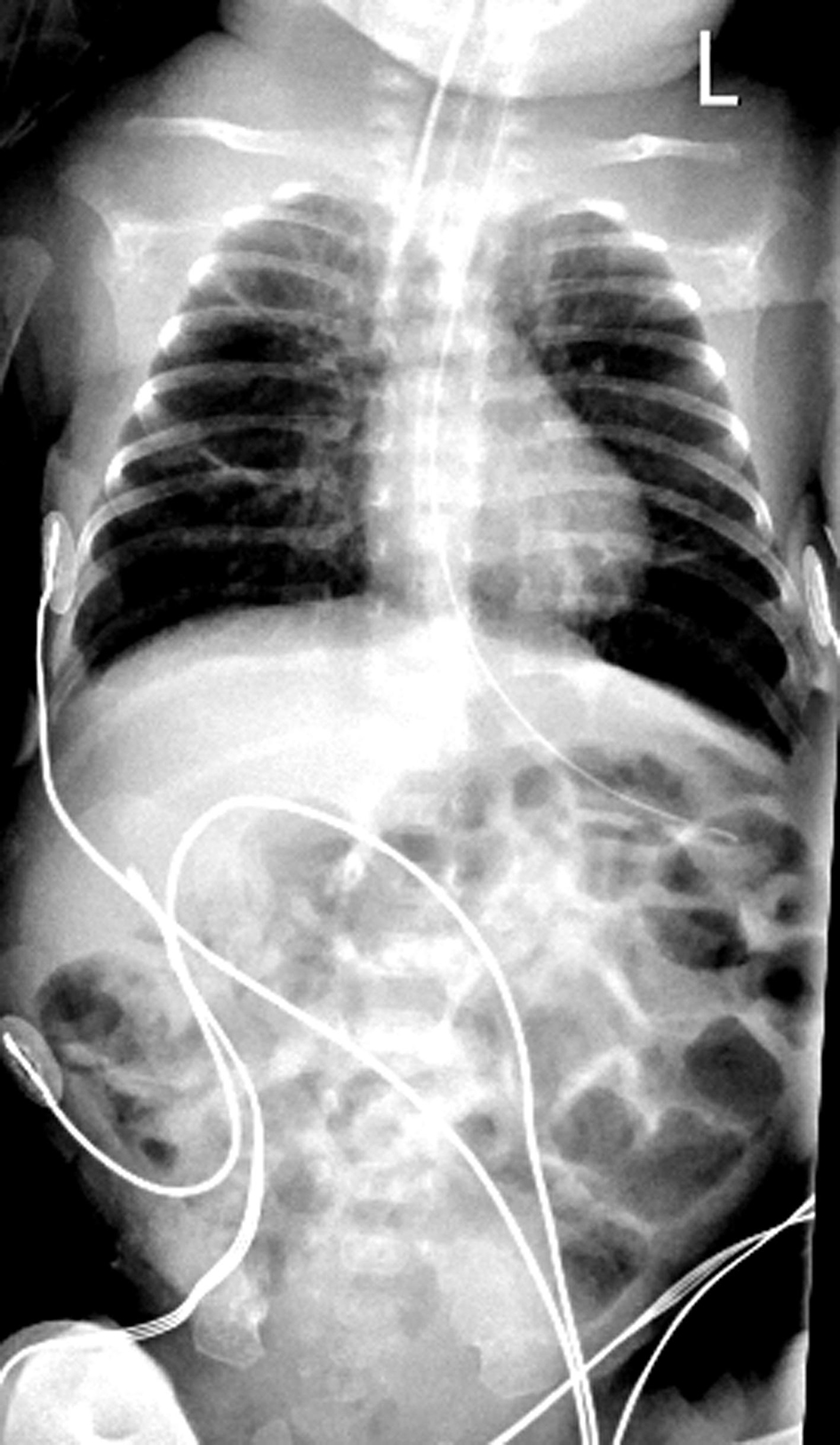
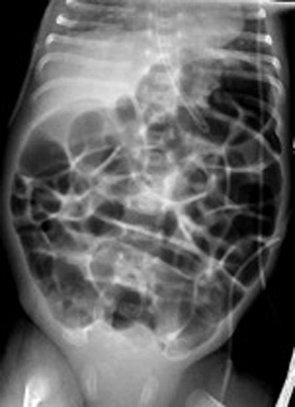
Figure 1. Chest and abdominal X-ray at 3 weeks of age, respiratory support with bubble CPAP of 8 cm of H2O and 6 L/min of gas flow in 25-30% FiO2 showing a normal distribution of the bowel gas pattern. CPAP: continuous positive airway pressure.
Figure 2.
Figure 2. Chest and abdominal X-rays. (a) At 5 weeks of age, was on respiratory support with bubble CPAP of 7 cm of H2O and 6 L/min of gas flow in 25-30 % FiO2 with the diagnosis of CPAP belly syndrome, immediately post-intubation. (b) Decubitus right body side up lateral shoot through X-ray film with no evidence of perforation. CPAP: continuous positive airway pressure.
Figure 3.
Figure 3. Chest and abdominal X-ray at 36 h post-intubation for CPAP belly syndrome showing reduced bowel gas pattern (decompression post-intubation, cessation of feeds) in comparison to Figure 2. CPAP: continuous positive airway pressure.
Figure 4.
Figure 4. Flow chart on the pathogenesis of CPAP belly syndrome. CPAP: continuous positive airway pressure.
Figure 5.
Figure 5. (a) Normal diaphragm and gastric contents. (b) Arrows indicate the downward pressure effects of CPAP on the diaphragm exerting pressure on the gastric contents, reducing gastric emptying time. CPAP: continuous positive airway pressure.
Figure 6.
Figure 6. Normal bowel wall ultrasound finding.