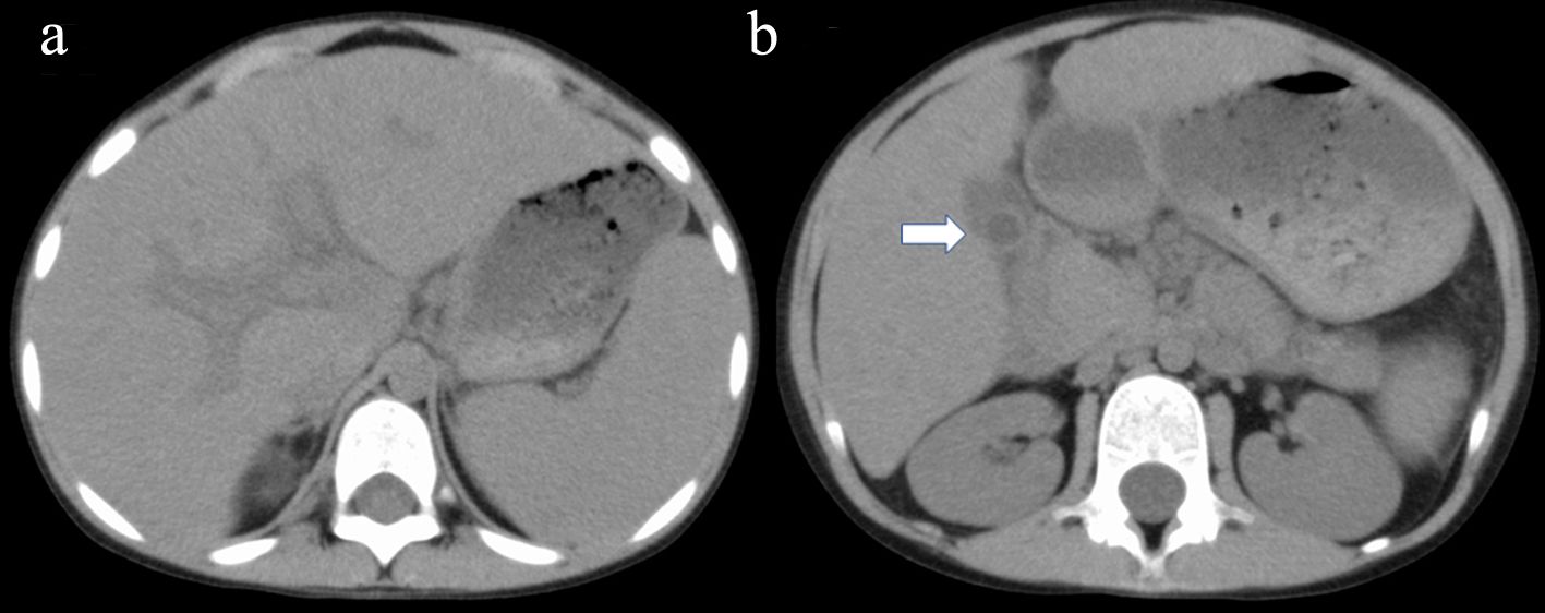
Figure 1. Abdominal axial CT shows hepato-splenomegaly associated with remarkable periportal collar sign (a) and edematous wall thickening of the gall bladder (b; arrow). CT: computed tomography.
| International Journal of Clinical Pediatrics, ISSN 1927-1255 print, 1927-1263 online, Open Access |
| Article copyright, the authors; Journal compilation copyright, Int J Clin Pediatr and Elmer Press Inc |
| Journal website http://www.theijcp.org |
Case Report
Volume 9, Number 2, June 2020, pages 50-54
Autoimmune Hepatitis With Severe Hypergammaglobulinemia and Eosinophilia in a Child
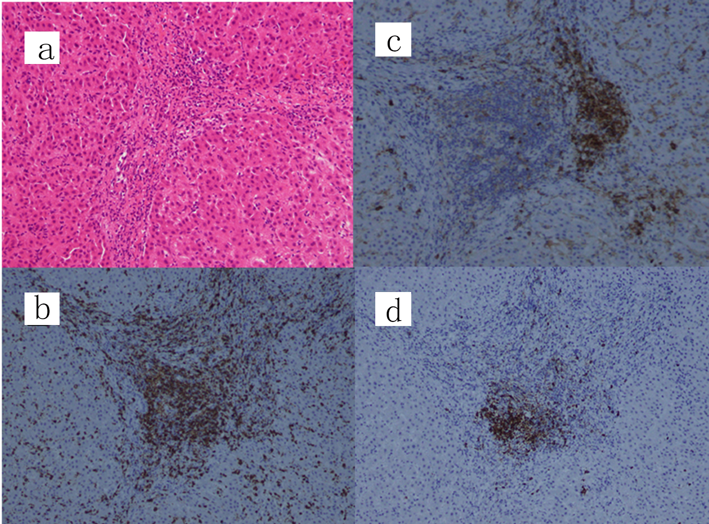
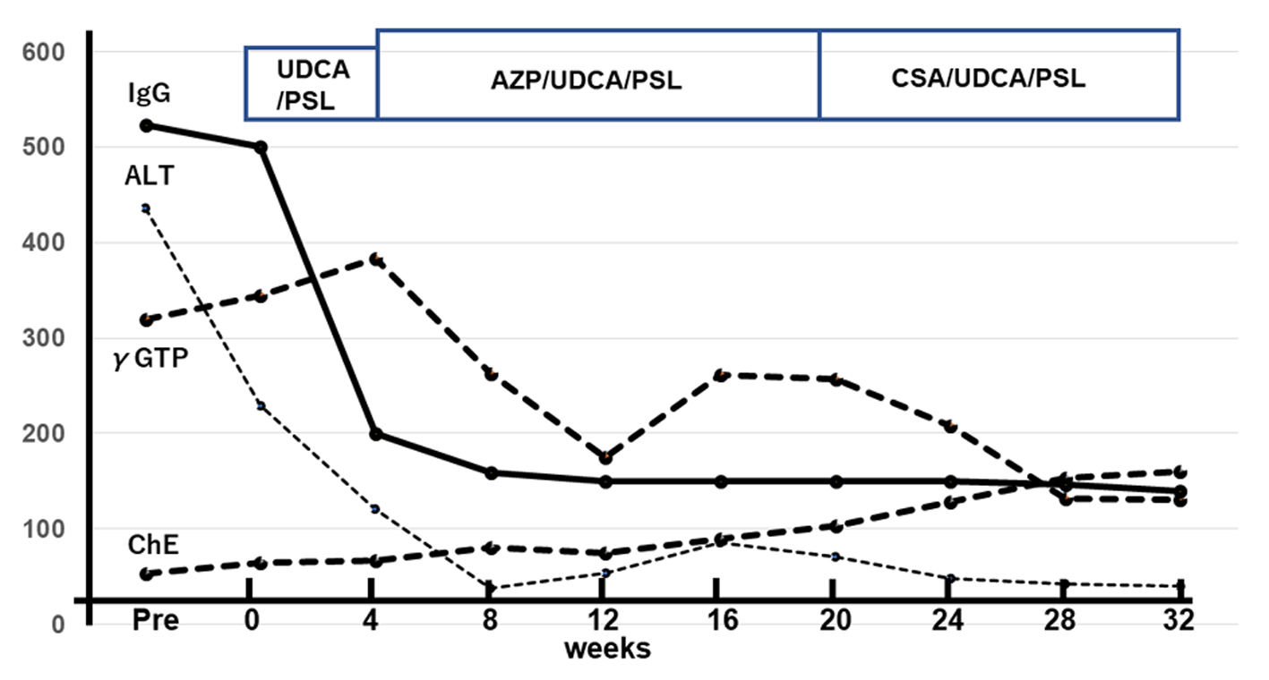
Figures