International Journal of Clinical Pediatrics, ISSN 1927-1255 print, 1927-1263 online, Open Access
Article copyright, the authors; Journal compilation copyright, Int J Clin Pediatr and Elmer Press Inc
Journal website https://www.theijcp.org

Case Report

Volume 11, Number 1, March 2022, pages 14-19


Recurrent Bilateral Pleural and Pericardial Effusions due to Tuberculosis in a Child With Doose Syndrome: A Case Report

Figures

Figure 1.
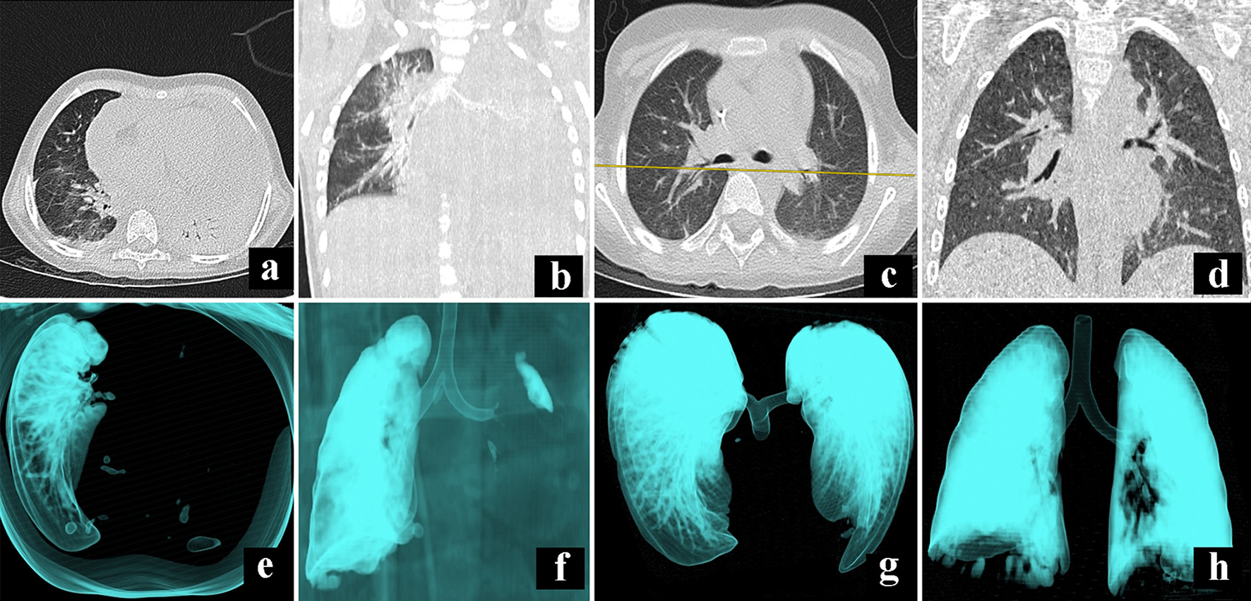
Figure 1. Comparative illustration of CT scan during admission (a, b, e, f) and first discharge (c, d, g, h). Panels (a) and (b) show a massive pleural effusion of the left lung that compromises all the segments; panels (e) and (f) show a 3D reconstruction to denote the occlusion of the left airway. Panels (c) and (d) show the absence of fluid in the pleural space after 18 days of treatment, while panels (g) and (h) show a full expansion of the lungs. CT: computed tomography.
Figure 2.
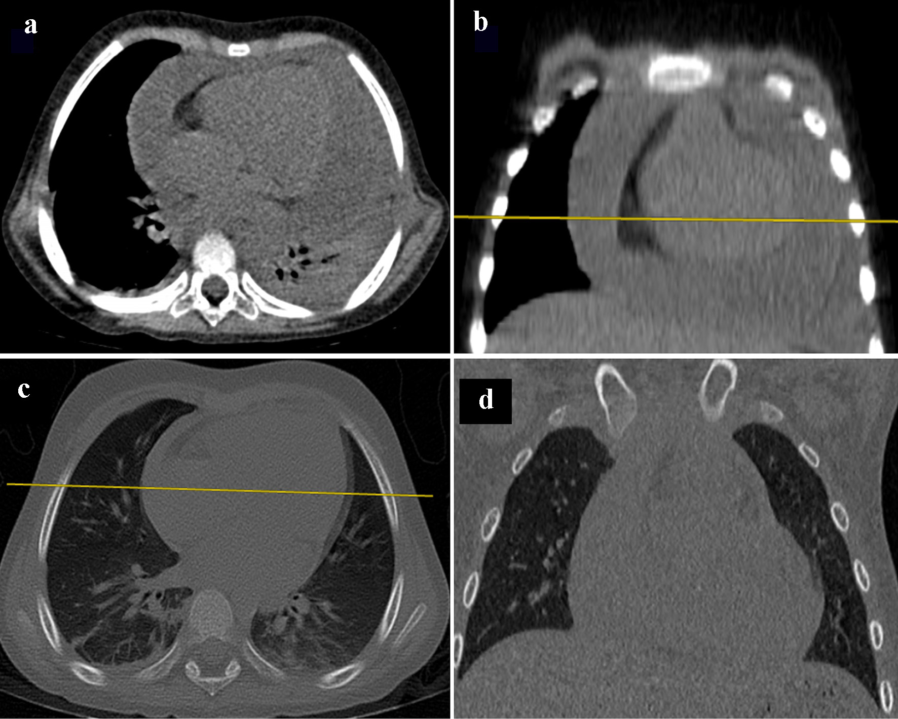
Figure 2. Pleural effusion image. In panels (a) and (b), CT scan of the mediastinum, with axial and coronal planes, show an increase of fluid in pericardial space. However, after treatment with diuretic therapy, comparative panels (c) and (d), the fluid collected in pleural space has diminished. CT: computed tomography.
Figure 3.
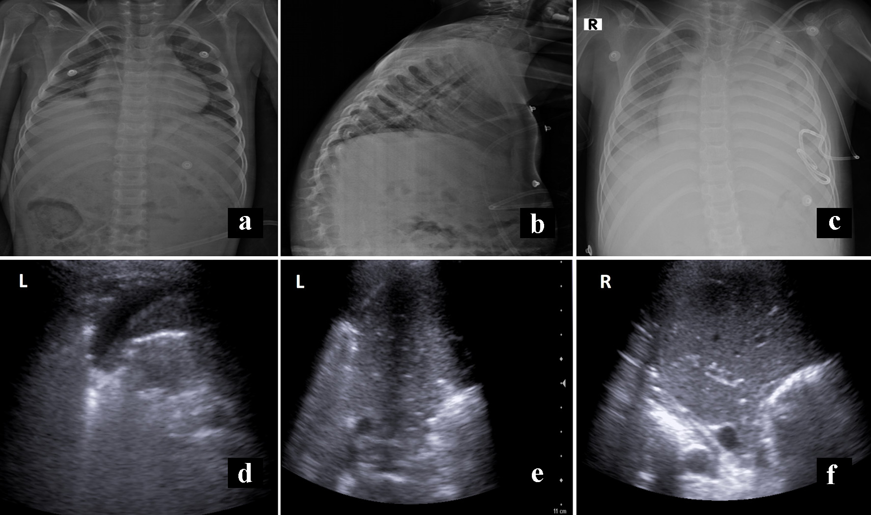
Figure 3. Panels (a) and (b) show the X-ray film before the first discharge without evidence of effusion. Panel (c) shows the initial film to second hospital readmission, with recurrent pleural effusion. Panels (d) and (e) correspond to sonography, previous to definitive discharge, with minimal fluid on the left side (L). In contrast, panel f shows lung parenchyma without fluid (R).

Tables

Table 1. Serum Analysis of Important Normochromic Normocytic Anemia
 
ValueValue
WBC: white blood cell; RBC: red blood cell; Hct: hematocrit; MCV: mean corpuscular volume; MCH: mean corpuscular hemoglobin; MCHC: mean corpuscular hemoglobin concentration.
WBC (× 103/mm3)3.82Urea (mg/dL)11.9
RBC334Creatinine (mg/dL)0.20
Hb (g/L)10.5Protein (mg/dL)5.4
Hct (%)32.3Albumin2.9
MCV (fL)96.7C-reactive protein12.1
MCH (pg)31.4Procalcitonin0.75
MCHC (g/dL)32.5Rheumatoid factor0
Platel count (× 103/mm3)89Immunoglobulin G545.60
Neutrophils (%)24Immunoglobulin M55.70
Lymphocytes (%)55Immunoglobulin A7.10
Monocytes (%)11Immunoglobulin E11.73
Eosinophils (%)7Complement component 3 (C3)143.6
Basophils (%)0Complement component 3 (C4)13.7
Lactic dehydrogenase185Antinuclear antibody test0.52
Glucose (mg/dL)89


Table 2. Pleural Fluid Characteristics
 
A high concentration of protein and leukocyte cells is shown. The negative reaction to Ziehl-Neelsen stain makes the diagnosis of tuberculosis infection hard to reach. LDH: lactate dehydrogenase.
Leukocyte count cells (/mm3)12,400pH8
Lymphocytes868Fungal cultureNegative
Neutrophyle10,664Glucose (mg/dL)77.5
LDH (U/L)155Ziehl-Neelsen stainingNegative
Count cells (/mm3)186Pleural effusion cultureNo growth
Protein (g/L)3.525Cytology reportMajority of lymphocytes.