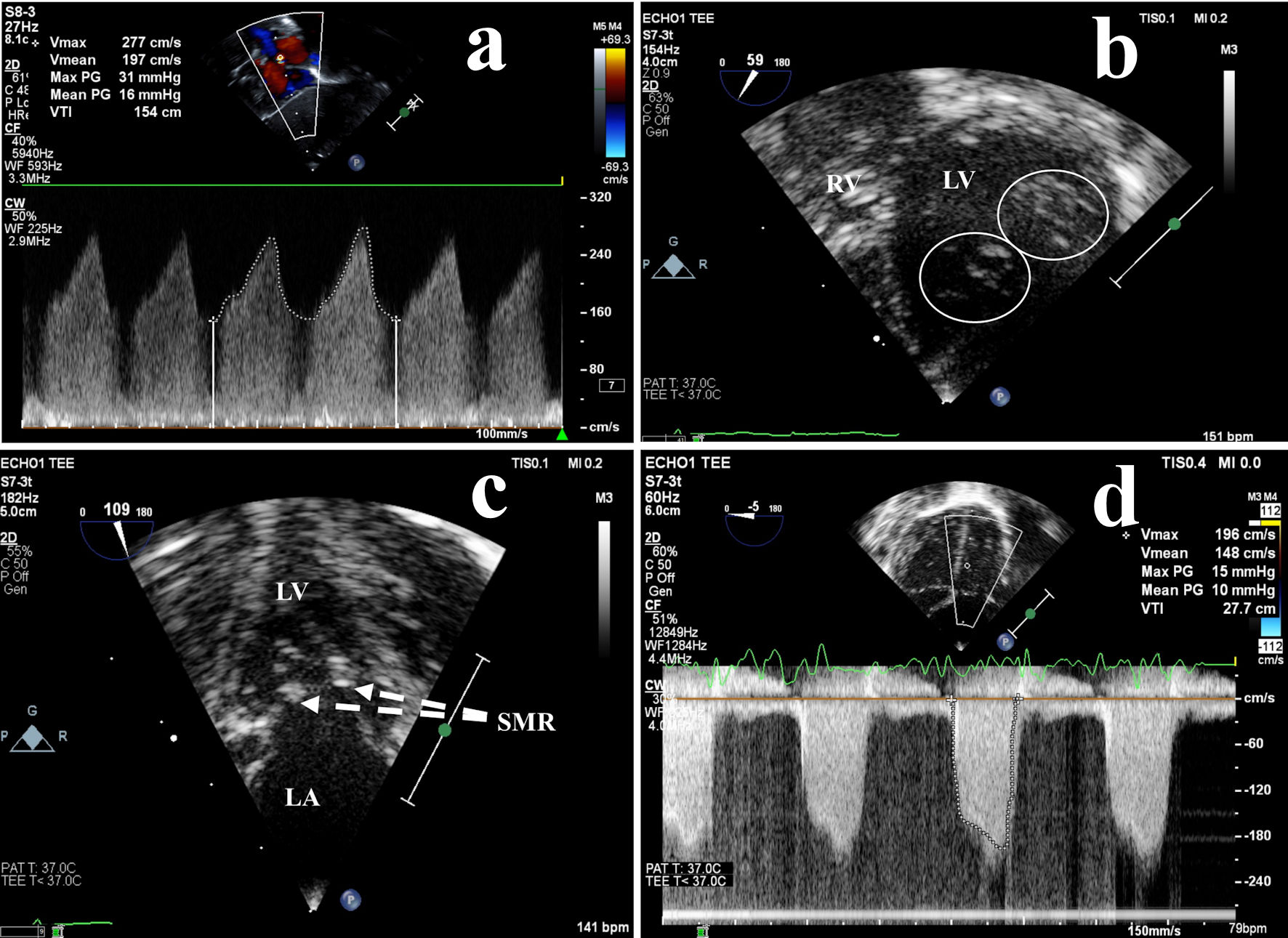
Figure 1. Preoperative echocardiography: left atrial hypertension due to obstructive mitral valve abnormalities. (a) High velocity left-to-right atrial shunt across the foramen ovale measured by transthoracic continuous wave Doppler, indicative of LA hypertension. Transesophageal echocardiography more clearly demonstrated (b) a double orifice mitral valve (circles) and (c) a supramitral ring (dashed lines), resulting in (d) moderate mitral valve inflow obstruction (stenosis). LA: left atrium; LV: left ventricle; PG: peak gradient; SMR: supramitral ring; RV: right ventricle; V: velocity.
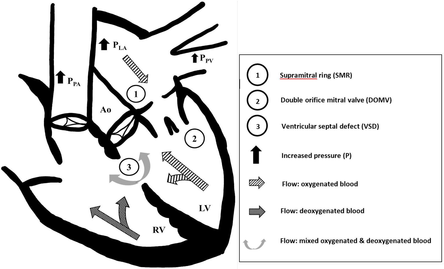
Figure 2. Constellation of acyanotic congenital heart disease leading to cyanotic physiology. We suspect that the (1) supramitral ring and (2) double orifice mitral valve obstructed left ventricular inflow, which increased left atrial (PLA) and pulmonary venous pressures (PPV), and caused bidirectional flow across the (3) ventricular septal defect (VSD). The left-to-right VSD flow further exacerbated left atrial and pulmonary venous pressures. Systemic desaturation was due to increasing elements of right-to-left VSD flow and pulmonary vascular congestion. Ao: aorta; LV: left ventricle; PLA: left atrium pressure; PPA: pulmonary artery pressure; PPV: pulmonary vein pressure; RV: right ventricle.