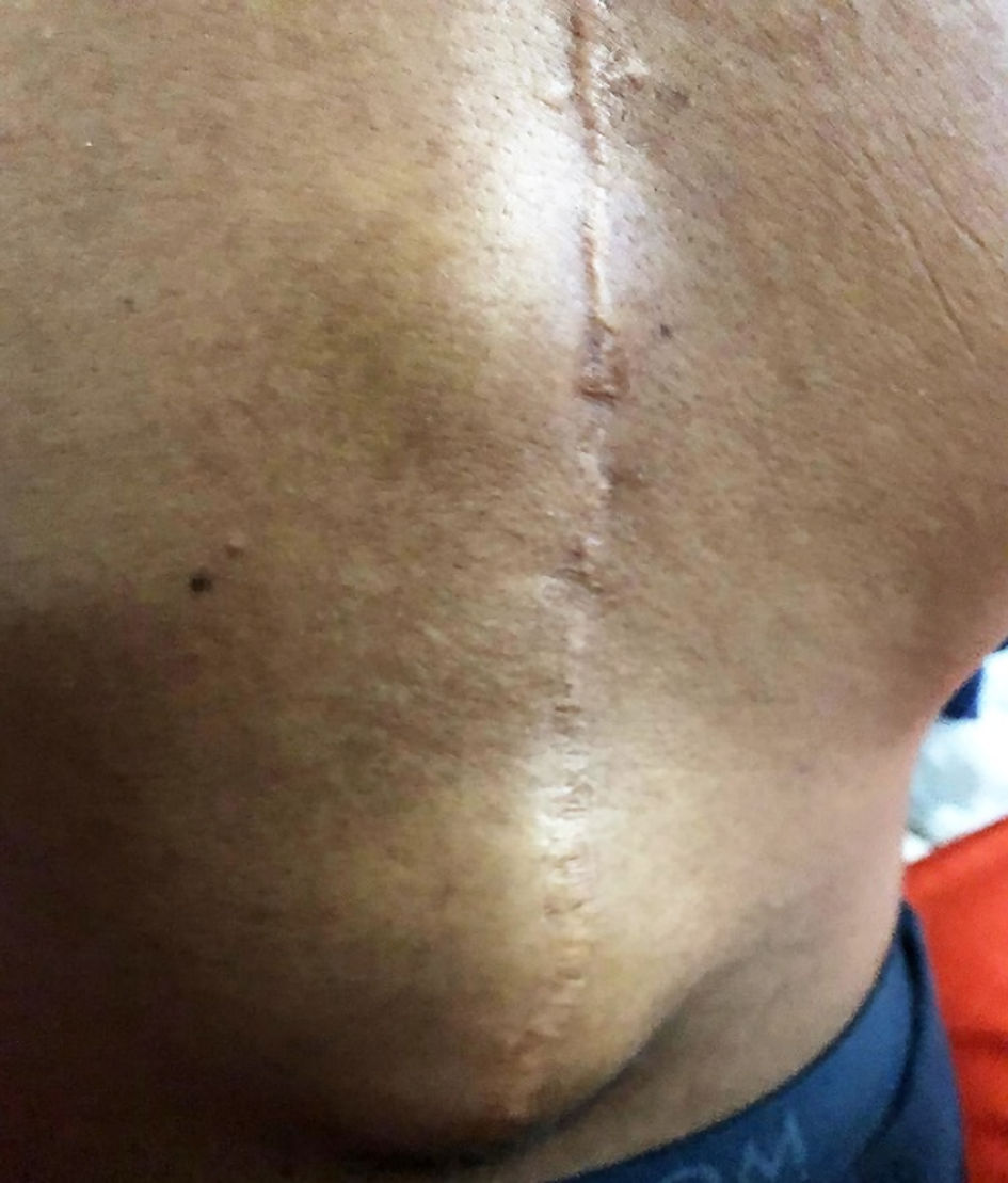
Figure 1. Photo of fluctuating subcutaneous swelling in the caudal part of the surgical scar around thoracic vertebra 12 and lumbar vertebra 1.
| International Journal of Clinical Pediatrics, ISSN 1927-1255 print, 1927-1263 online, Open Access |
| Article copyright, the authors; Journal compilation copyright, Int J Clin Pediatr and Elmer Press Inc |
| Journal website https://www.theijcp.org |
Case Report
Volume 11, Number 2, June 2022, pages 60-65
Salmonella Osteomyelitis in Two Immunocompetent Children
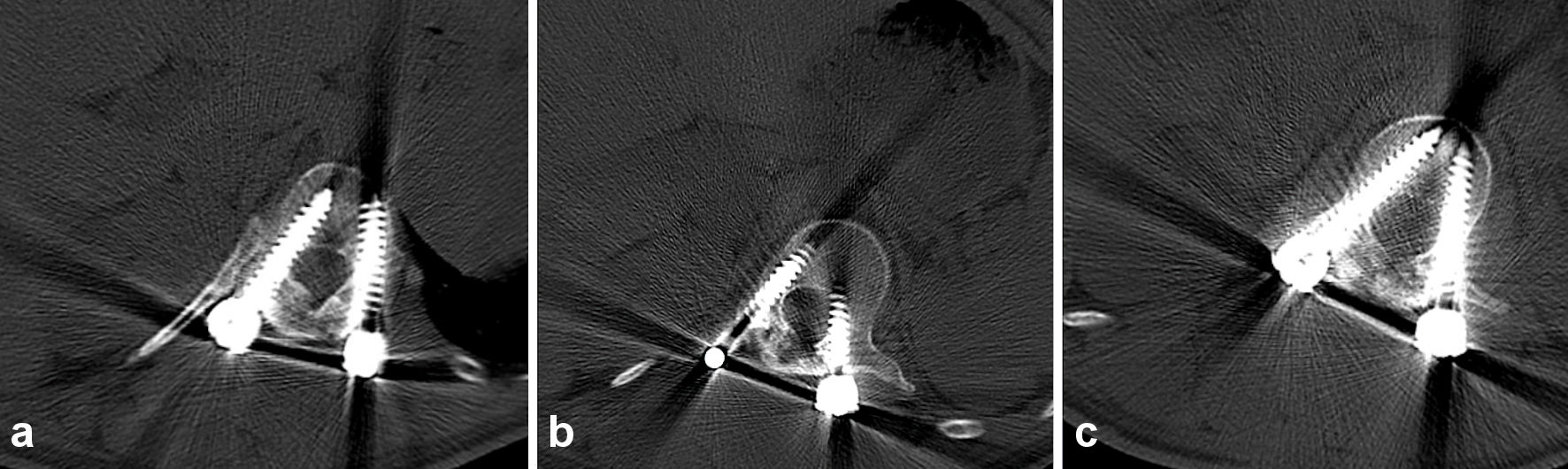
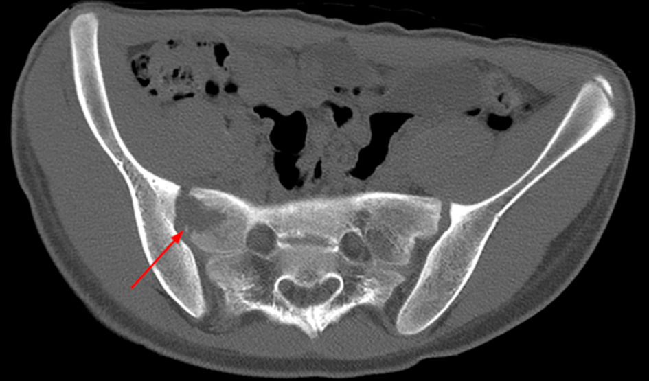
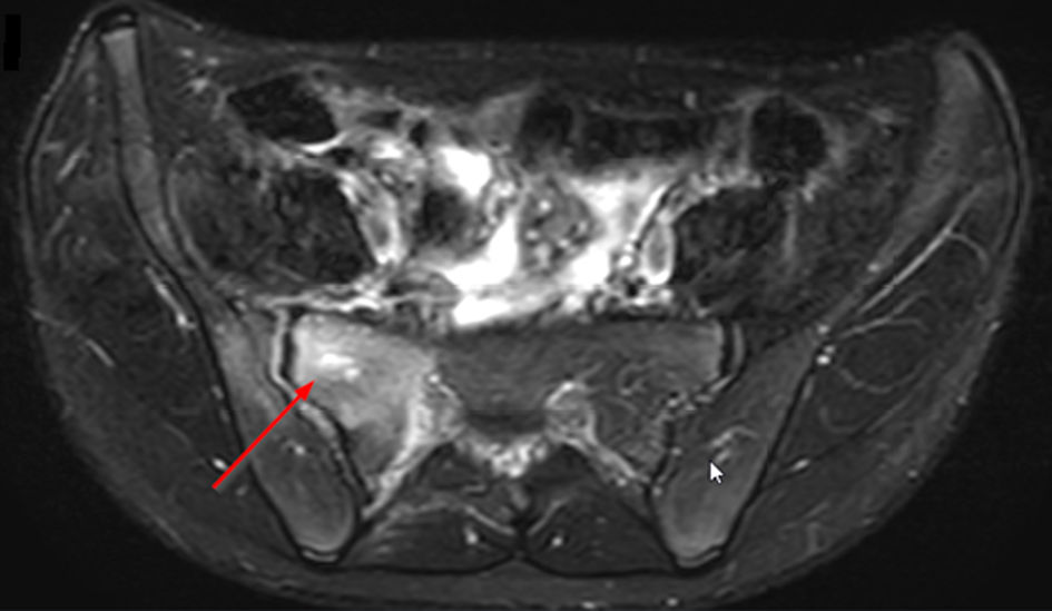
Figures