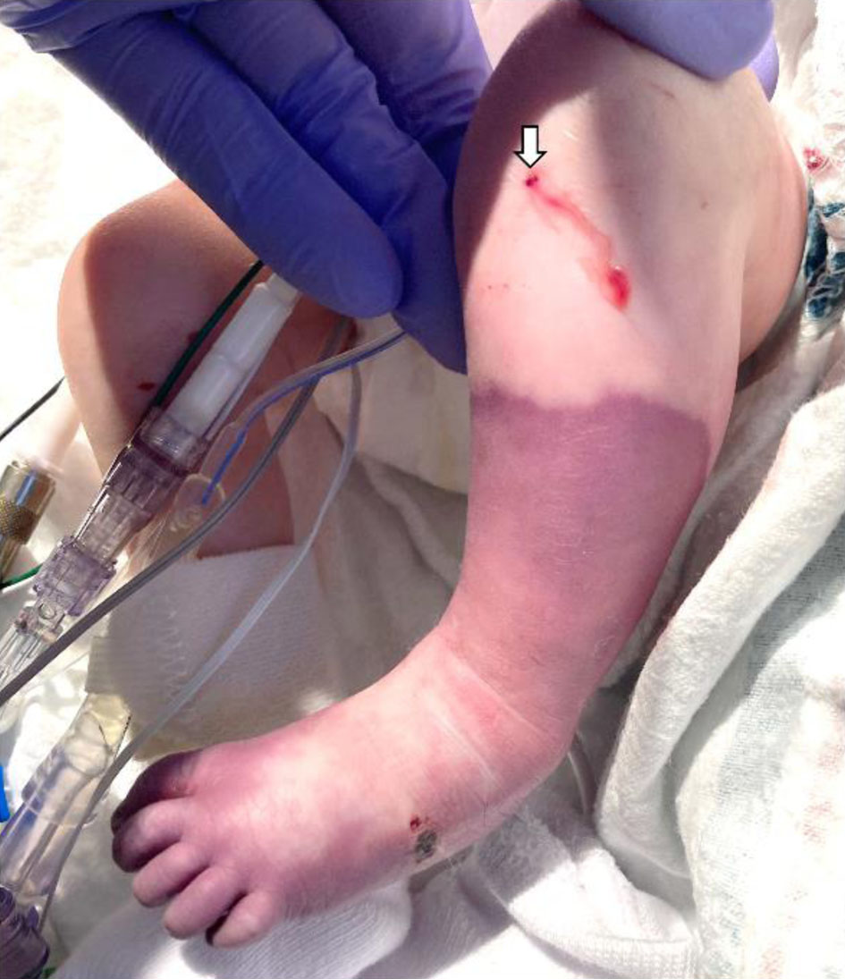
Figure 1. Following the resuscitation, there was progressive decreased perfusion and cyanosis of the left lower extremity with significant color change from the mid-tibia distally to the foot. The intraosseous needle insertion site is noted (white arrow).
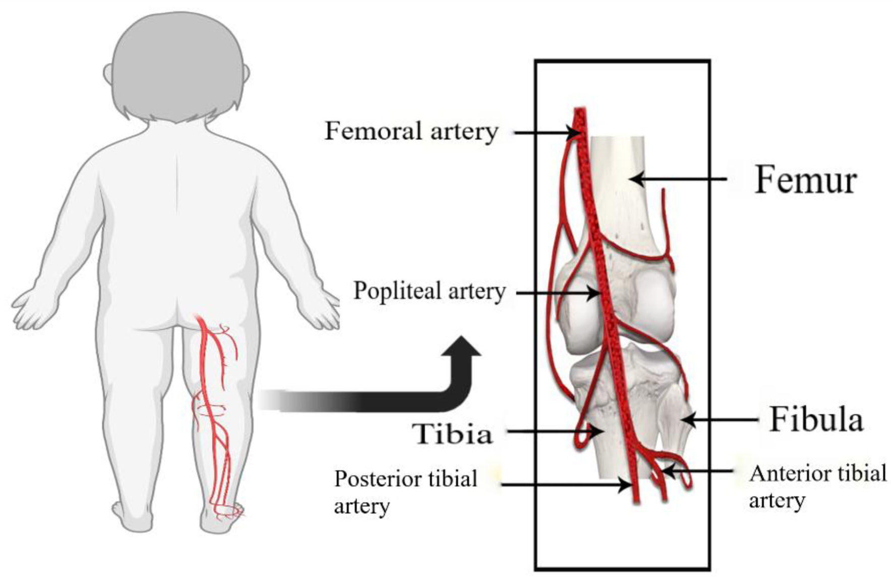
Figure 3. Diagram showing the course of the popliteal artery. The femoral artery, a continuation of the external iliac artery travels through the adductor canal on the medial side of the femur above the medial epicondyle. The adductor canal, an opening in the adductor magnus muscle hiatus, is located at the junction of the middle and lower thirds of the thigh. The artery is anterior to the femur in the femoral canal, but then travels anteromedially, and crosses behind the medial epicondyle of the femur into the popliteal fossa. In the popliteal fossa, it becomes the popliteal artery and travels posterior to the tibia.