International Journal of Clinical Pediatrics, ISSN 1927-1255 print, 1927-1263 online, Open Access
Article copyright, the authors; Journal compilation copyright, Int J Clin Pediatr and Elmer Press Inc
Journal website http://www.theijcp.org

Case Report

Volume 1, Number 4-5, October 2012, pages 124-128


Mumps-virus-associated Clinically Mild Encephalopathy With a Reversible Splenial Lesion

Figures

Figure 1.
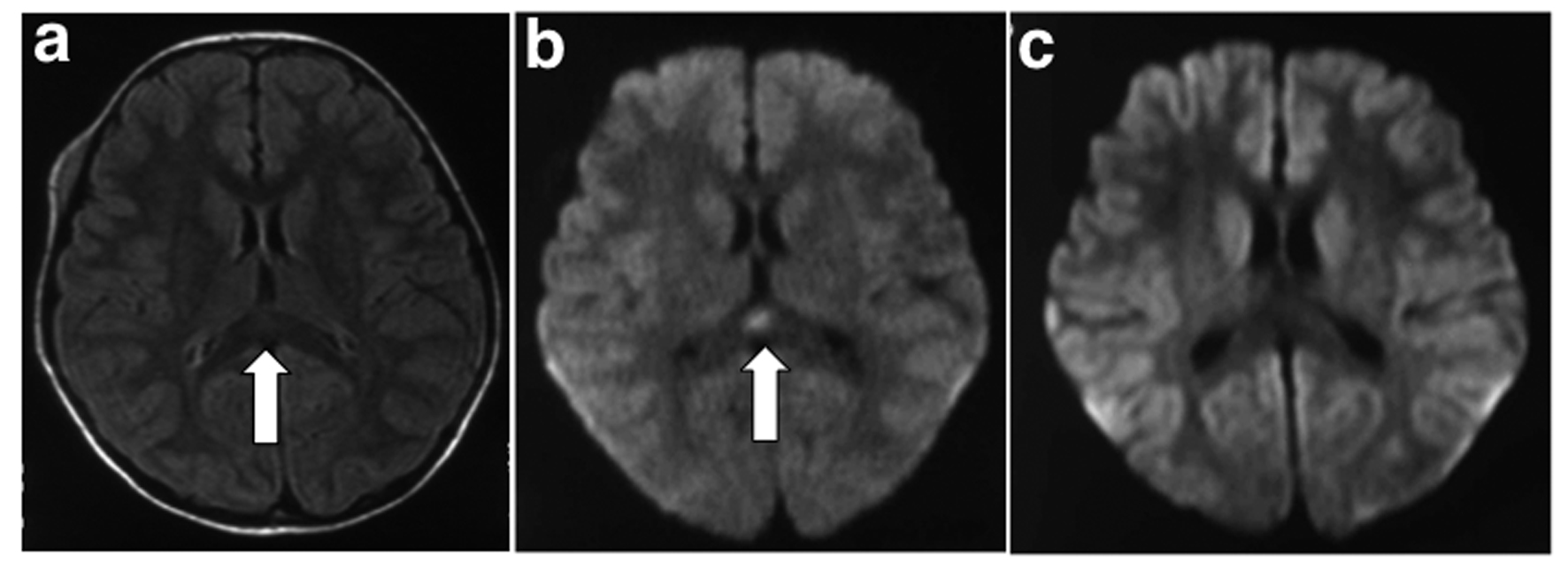
Figure 1. Brain magnetic resonance imaging. a: Fluid-attenuated inversion recovery image on the second day of admission (third day from onset) showed a slightly hyperintense lesion in the splenium of the corpus callosum (arrow); b: Diffusion-weighted image on the second day of admission (third day from onset) showing a focal high-intensity lesion in the splenium of the corpus callosum (arrow); c: Diffusion-weighted image on eighth day of admission (ninth day from onset) showing complete resolution of the lesion.
Figure 2.
Figure 2. EEG in awake patient (third day from onset) showing a global diffuse, high-voltage, slow wave about 3 - 4 Hz in the particularly marked occipital area. Calibration: 100 µV, 1 s.

Table

Table 1. Previously Published Reports of MERS Associated With Mumps Virus Infection
 
ReferencesPatient (age, sex)CNS manifestationCNS diagnosisCSF CC (/µL)EEGTherapyOutcome
MERS, clinically mild encephalitis/encephalopathy with a reversible splenial lesion; CNS, central nervous system; CSF cerebrospinal fluid; CC, cell count; EEG, electroencephalogram; M, male; F, female; LOC, level of consciousness; BA, basic activity; IVIgG, intravenous immunoglobin; NE, not explained; DIV, drip intravenous isotonic fluid infusion.
Tada et al, 2004 [2]7 years, MDecreased LOCMeningoencephalitisPleocytosisSlow BAIVIgGHealthy
Tada et al, 2004 [2]8 years, MDecreased LOC, seizureMeningoencephalitis119NormalNEHealthy
Matsumoto et al, 2009 [4]5 years, FDecreased LOCMeningoencephalitis132Slow BADIVHealthy
This case4 years, FDecreased LOC, seizureEncephalopathy1Slow BADIV, D-mannitolHealthy