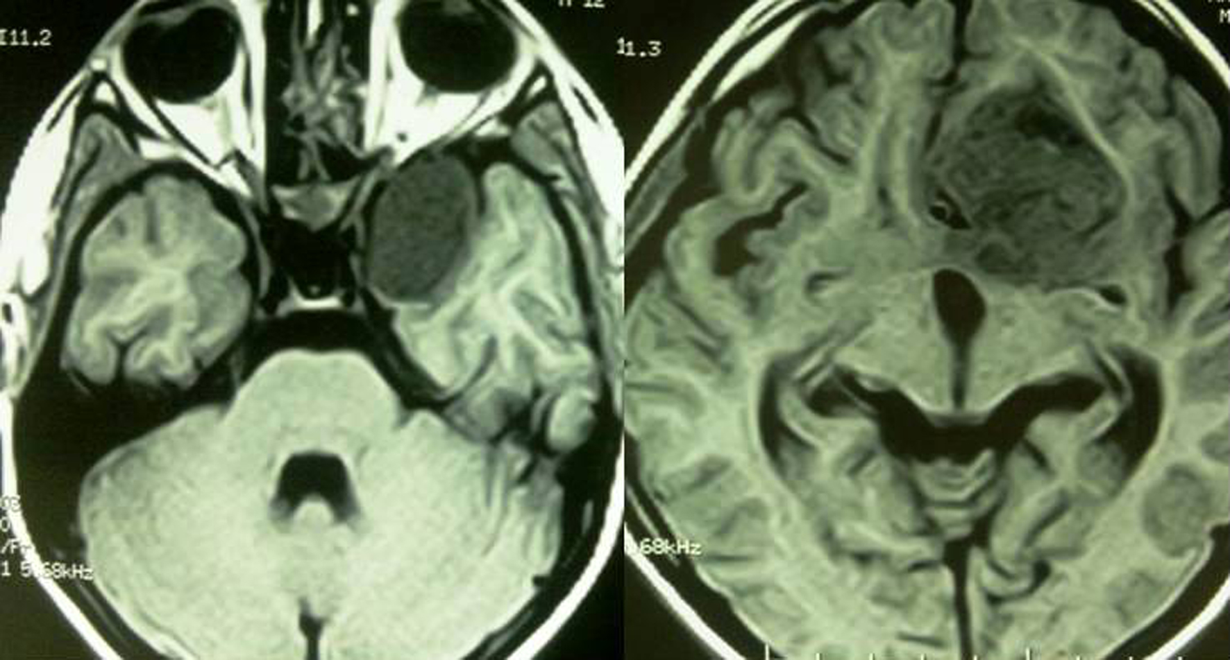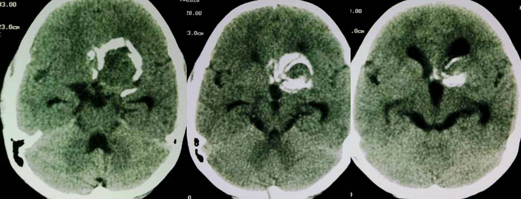
Figure 1. Axial non contrast CT images reveal a densely calcified soft tissue density lesion in the suprasellar region with associated dilatation of third and lateral ventricles.
| International Journal of Clinical Pediatrics, ISSN 1927-1255 print, 1927-1263 online, Open Access |
| Article copyright, the authors; Journal compilation copyright, Int J Clin Pediatr and Elmer Press Inc |
| Journal website http://www.theijcp.org |
Case Report
Volume 1, Number 4-5, October 2012, pages 129-132
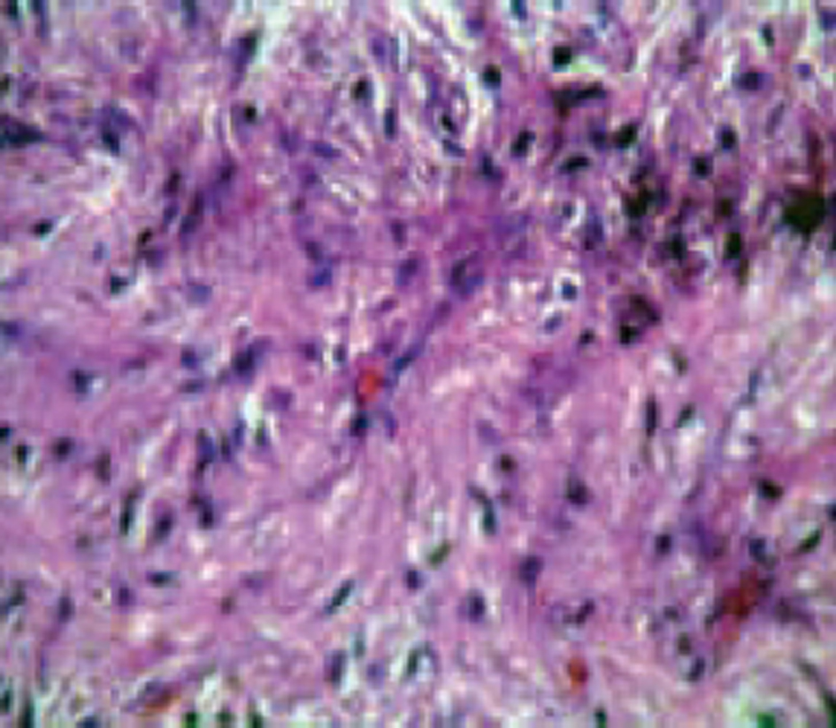
Densely Calcified Pilocytic Astrocytoma in the Sellar/Suprasellar Region
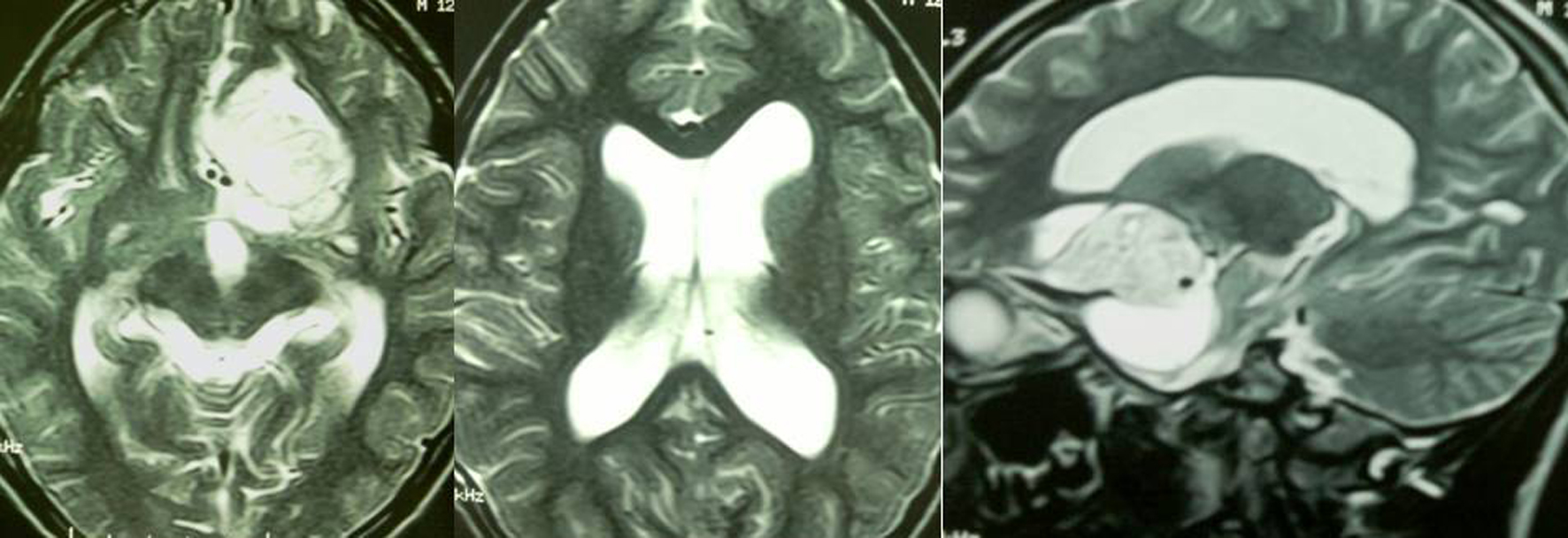
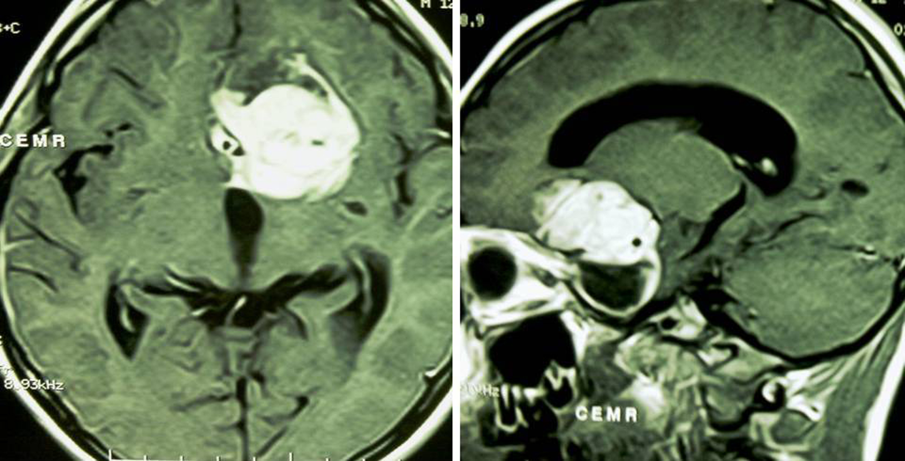
Figures