

| International Journal of Clinical Pediatrics, ISSN 1927-1255 print, 1927-1263 online, Open Access |
| Article copyright, the authors; Journal compilation copyright, Int J Clin Pediatr and Elmer Press Inc |
| Journal website http://www.theijcp.org |
Case Report
Volume 4, Number 2-3, October 2015, pages 158-161
Clinical Significance of Bacille Calmette-Guerin Inoculation Site Erythema in Incomplete Kawasaki Disease
Ahmad Jaafara, c, Eman Al-Hashemib
aDepartment of Pediatrics, Mubarak Al-Kabeer Hospital, Kuwait
bDepartment of Medicine, Mubarak Al-Kabeer Hospital, Kuwait
cCorresponding Author: Ahmad Jaafar, Department of Pediatrics, Mubarak Al-Kabeer Hospital, Kuwait
Manuscript accepted for publication July 09, 2015
Short title: BCG Site Erythema and Kawasaki Disease
doi: http://dx.doi.org/10.14740/ijcp211w
| Abstract | ▴Top |
Kawasaki disease is an acute systemic vasculitis occurring mainly in infants and children. It is considered a clinical diagnosis, diagnosed by a set of clinical criteria. When the needed criteria to make the diagnosis are not met, a diagnostic dilemma occurs that might contribute to a delay in the diagnosis and treatment, hence increasing the risk of its most feared complication, namely coronary artery aneurysm. This is particularly true in infants, who tend to have the incomplete as well as complicated form of the disease. This is why having additional specific and reliable clinical clues to the diagnosis can be of great value. We present an 8-month-old boy, who presented with a 4-day history of fever and fulfilled three out of the five clinical criteria needed to make the diagnosis of Kawasaki disease; however, due to the presence of Bacille Calmette-Guerin (BCG) inoculation site erythema, the delay in the diagnosis and treatment was avoided. The medical literature lacks sufficient research regarding the usefulness of this sign in Kawasaki disease. This study aims to emphasize on the importance of the erythema around the BCG inoculation site in the diagnosis of the challenging cases of incomplete Kawasaki disease, mainly in infants.
Keywords: Kawasaki disease; Incomplete; Vasculitis; BCG; Erythema; Infant
| Introduction | ▴Top |
Kawasaki disease, first described by Dr. Tomisaku Kawasaki in Japan in 1967, is an acute febrile vasculitis affecting medium-sized arteries, mainly in children less than 5 years of age [1, 2]. Kawasaki disease has been reported in various countries from all around the world but mostly in Asia [3]. Its etiology still remains unknown; however, epidemiological data suggest that an immune response to an infectious agent, occurring in genetically susceptible people, might be the initial trigger [4-6]. There is not a specific diagnostic test to diagnose Kawasaki disease as it is primarily a clinical diagnosis based on the presence of a specific set of clinical criteria. These criteria, which are approved by the American Heart Association (AHA), are the presence of fever for at least 5 days as well as non-purulent conjunctivitis, rash, cervical lymphadenopathy, and changes in extremities [7]. It is usually self-limiting; however, early treatment is recommended, as delayed treatment is considered a risk factor for the development of its most feared complication, namely coronary artery aneurysm [7, 8]. Coronary artery disease, most specifically coronary artery aneurysm, occurs in about 20% of untreated patients, making Kawasaki disease a more common cause of acquired cardiac disease in childhood than rheumatic fever in Japan and the United States [1]. In infancy, Kawasaki disease has a specific consideration in that it tends to be incomplete, which means that it tends not to fulfill all the clinical criteria needed to make the diagnosis, and thus creates a diagnostic dilemma [7]. Similarly, infancy is considered a risk factor for developing a more complicated disease [7]. This is why early diagnosis and treatment are of particular significance in infancy. From this point of view, having additional reliable clinical clues to the diagnosis can be crucial. One of such clues that is reported and discussed in this case report is the erythema around the BCG inoculation site.
The study aims to emphasize on the importance of the erythema around the BCG inoculation site in the early diagnosis of Kawasaki disease, particularly incomplete Kawasaki disease.
| Case Report | ▴Top |
We present an 8-month-old Kuwaiti boy, previously healthy, presented with a 4-day history of fever, which was documented by an ear thermometer, reaching a maximum of 39.4 °C, intermittent in nature and associated with irritability, decreased feeding and decreased activity. The fever was responding better to NSAIDs (ibuprofen) than paracetamol. On the third day of fever, the patient developed bilateral red eyes, red lips and redness (erythema) around the BCG inoculation site. The patient did not develop other skin rash or changes in the hands or feet.
There is no history of cough, difficulty in breathing, nasal blockage, rhinorrhea or sneezing. Additionally, there is no history of rigors, abnormal movements, convulsions, altered level of consciousness, change of bowel habits, vomiting, change in the color, odor or amount of urine or stool, contact with sick people or recent travel history.
The child has been growing and developing normally, is vaccinated up-to-date, on a healthy mixed diet, with unremarkable past medical, surgical or perinatal histories. The parents are non-consanguineous. The patient has a healthy older brother, and the family history is unremarkable for any genetic disease, autoimmune disease or malignancy.
On examination, the child looked irritable, in mild dehydration with no respiratory distress. The vital signs were as follows: heart rate 120/min, respiratory rate 30/min, blood pressure 90/70 mm Hg, body temperature 37.8 °C, and oxygen saturation 99% on room air. Examination of the eyes showed evidence of bilateral non-purulent conjunctivitis with no exudate, pallor or jaundice. The lips were red with minimal cracking. The tongue and oropharynx were normal. Neck examination revealed cervical lymphadenopathy, about 1 cm in diameter, mainly on the right side. The axillary and inguinal lymph nodes were not enlarged. Skin examination was unremarkable, apart from a 2-cm erythema surrounding the BCG inoculation site (Fig. 1). The examination of the chest, heart, abdomen, nervous system, ears and back were unremarkable. Examination of the genitals showed evidence of right-sided hydrocele.
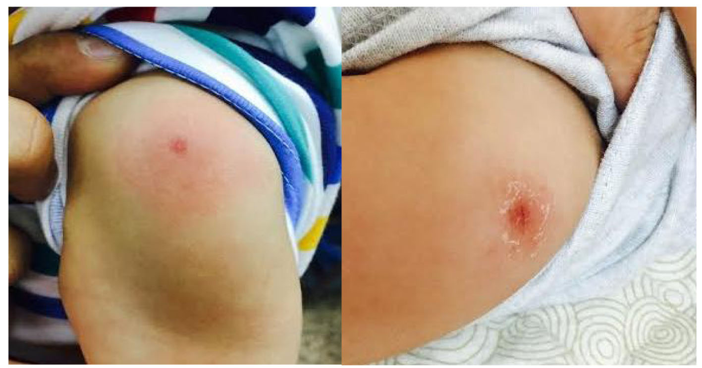 Click for large image | Figure 1. Left: the disappearance of the erythema around the BCG inoculation site 36 h following the administration of IVIG. Right: the erythema around the BCG inoculation site on admission (before the administration of IVIG). |
Investigations obtained initially include blood and urine work-up, electrocardiograph (ECG) as well as chest X-ray (CXR). The blood and urine work-up results are as shown in Tables 1 and 2, respectively. ECG showed a normal sinus rhythm and the CXR showed a normal cardiac silhouette with no evidence of cardiac or lung disease.
 Click to view | Table 1. Blood Work-Up Results of the 8-Month-Old Boy in the Current Case Report |
Click to view | Table 2. Urine Work-Up Results of the 8-Month-Old Boy in the Current Case Report |
The patient was admitted to the hospital, started on intravenous hydration, IV antibiotic and antipyretic measures (paracetamol, ibuprofen and cold sponging) for fever. Our initial impression was Kawasaki disease, but due to the fact that not all of the criteria of the disease were met, particularly the duration of the fever, an infection was suspected and this is why a partial septic screen was taken and an IV antibiotic was started; however, a rheumatologist was consulted, who strongly suspected Kawasaki disease and decided to start the patient on intravenous immunoglobulin (IVIG) (2 mg/kg over 12 h) after a dose of intravenous hydrocortisone as well as oral aspirin (5 mg/kg/day). After about 16 h from the start of IVIG, the fever subsided and the conjunctivitis, red lips and BCG inoculation site erythema started to improve. After about 36 h, the previous features, specifically the erythema around the BCG inoculation site disappeared completely (Fig. 1). Once the dose of IVIG finished and the fever subsided, the patient was shifted to the pediatric echocardiograph department for a proper 2D departmental echocardiograph. The echocardiograph showed evidence of right coronary artery mild ectasia (1.9 mm) (normal is up to 1.7 mm) (Fig. 2). Following the echocardiograph, the patient was discharged home on daily oral aspirin (5 mg/kg/day) and a follow-up echocardiograph after 14 days.
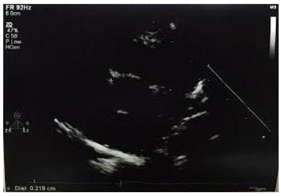 Click for large image | Figure 2. Two-dimensional departmental echocardiograph showing the mild dilatation of the right coronary artery (ectasia). |
| Discussion | ▴Top |
Kawasaki disease is an acute febrile systemic vasculitis of the medium-sized arteries of an unknown etiology, occurring almost exclusively in children. It is primarily a clinical diagnosis, diagnosed by a set of clinical criteria. To diagnose this disease according to AHA, there should be at least a 5-day history of documented fever as well as four out of five clinical features, which are changes in extremities (swelling, erythema or peeling), polymorphous rash, cervical lymphadenopathy (usually unilateral, > 1.5 cm in diameter), non-purulent conjunctivitis and changes of the lips, oral cavity and oropharynx (red lips with or without cracking, red tongue (strawberry tongue) and congested oropharynx) [7]. When the fever lasts less than 5 days, a diagnostic dilemma occurs. According to AHA guidelines, a 5-day history of fever is needed to make the diagnosis [7]. On the other hand, from the Japanese Circulation Society guidelines, Kawasaki disease can be diagnosed even before the completion of 5 days of fever [9]. Concerning the five other clinical criteria, AHA emphasizes on the importance of having four out of five criteria in order to diagnose the disease, and when less than four criteria are met, the patient could have what is called incomplete Kawasaki disease. This might cause a dilemma and contribute to the delay in making the diagnosis, and hence giving the treatment [10]. This form of the disease is more common in infants [7]. From what mentioned above, one can conclude that diagnosing a case of Kawasaki disease, particularly during infancy, can be challenging. This is seen in our patient who presented with a 4-day history of fever and three out of the five clinical criteria with a normal initial laboratory work-up.
The most feared complication of Kawasaki disease is coronary artery disease, especially coronary artery aneurysm, which affects about 20% of untreated patient [1]. Early in the course of the illness, the aneurysm may not be present; however, evidence of coronary artery affection like ectasia might be present. This complication is more common in infants than older children [7]. Additionally, it is more common in patients in whom the diagnosis and treatment were delayed [8]. Additionally, the risk of developing coronary artery aneurysm is similar between the incomplete and classical forms of Kawasaki disease [11]. When referring to our case, we can notice that despite the fact that the presentation was not classical for Kawasaki disease concerning the duration of the fever and the number of fulfilled criteria, the patient already started to have coronary artery affection. Fortunately, there was no delay in making the diagnosis and giving the treatment.
As mentioned previously, one can safely reach to the conclusion that infants are at particular risk of developing the more complicated as well as incomplete form of the disease, which makes finding additional signs in them for early diagnosis a priority [12]. This is why the search for more specific clinical clues to aid in the diagnosis in equivocal cases is crucial, particularly during infancy.
Erythema around the BCG inoculation site in our patient was of great significance as it helped in the early diagnosis and the prevention of a possible delay in giving the treatment. This sign is encountered in a number of cases of Kawasaki disease, and there is a limited medical literature about its usefulness in the disease. Although, it is not considered a criterion in the diagnosis of Kawasaki disease, it has been reported to be a specific sign of the disease [13, 14]. This sign is of particular importance in areas where the BCG vaccination is universal like in Kuwait. The mechanism by which the erythema occurs remains controversial. It has been hypothesized that there is cross-reactivity between the mycobacterial heat shock protein (HSP) 65 and the human homologue HSP63, a mitochondrial protein. The detection of strong antibody and cellular reactivity against both the synthetic peptides of the mycobacterial HSP65 and the HSP63 in the blood of children with Kawasaki disease goes in line with this hypothesis [15].
Conclusion
Early diagnosis and treatment of Kawasaki disease are associated with a better overall outcome. Since Kawasaki disease is primarily a clinical diagnosis based on the presence of a set of clinical criteria, the lack of sufficient clinical criteria makes early diagnosis and hence early treatment a challenge. This is particularly true in infants who tend to have the incomplete as well as complicated form of the disease. Erythema around the BCG inoculation site can help in the early diagnosis of challenging cases of incomplete Kawasaki disease, as it is a specific and reliable clinical clue to the diagnosis.
Competing Interests
The authors declare that they have no competing interests.
| References | ▴Top |
- Rowley AH, Shulman ST. Kawasaki disease. In Nelsons Text Book of Pediatrics. 19th ed. Behrman RE, Kliegman RM, Jenson HB,eds. Elsevier. 2011:823-826.
- Burns JC, Glode MP. Kawasaki syndrome. Lancet. 2004;364(9433):533-544.
doi - Nakamura Y, Yanagawa H. The worldwide epidemiology of Kawasaki disease. Prog Pediatr Cardiol. 2004;19:99-108.
doi - Rowley AH. Kawasaki disease. In: Gershon AA, Hotes PJ, Katz SL, et al. Krugmans Infectious Diseases of Children. 11th ed. Philadelphia, PA: Mosby Inc. 2004;323-335.
pubmed - Wang CL, Wu YT, Liu CA, Kuo HC, Yang KD. Kawasaki disease: infection, immunity and genetics. Pediatr Infect Dis J. 2005;24(11):998-1004.
doi pubmed - Yeung RS. Kawasaki disease: update on pathogenesis. Curr Opin Rheumatol. 2010;22(5):551-560.
doi pubmed - Newburger JW, Takahashi M, Gerber MA, Gewitz MH, Tani LY, Burns JC, Shulman ST, et al. Diagnosis, treatment, and long-term management of Kawasaki disease: a statement for health professionals from the Committee on Rheumatic Fever, Endocarditis and Kawasaki Disease, Council on Cardiovascular Disease in the Young, American Heart Association. Circulation. 2004;110(17):2747-2771.
doi pubmed - Palinkas LA, Wilder MS, Kao AS, et al. Social and cultural risk factors for coronary artery aneurysms following Kawasaki syndrome. Pediatr Res. 2003;53:326A.
- Ogata S, Bando Y, Kimura S, Ando H, Nakahata Y, Ogihara Y, Kaneko T, et al. The strategy of immune globulin resistant Kawasaki disease: a comparative study of additional immune globulin and steroid pulse therapy. J Cardiol. 2009;53(1):15-19.
doi pubmed - Witt MT, Minich LL, Bohnsack JF, Young PC. Kawasaki disease: more patients are being diagnosed who do not meet American Heart Association criteria. Pediatrics. 1999;104(1):e10.
doi pubmed - Sonobe T, Kiyosawa N, Tsuchiya K, Aso S, Imada Y, Imai Y, Yashiro M, et al. Prevalence of coronary artery abnormality in incomplete Kawasaki disease. Pediatr Int. 2007;49(4):421-426.
doi pubmed - Rosenfeld EA, Corydon KE, Shulman ST. Kawasaki disease in infants less than one year of age. J Pediatr. 1995;126(4):524-529.
doi - Plantin P, Blayo M, Dupre D, et al. BCG reactivation: A rare but specific sign of Kawasaki Disease. Press Med. 1998;27(15):716.
pubmed - Takayama J, Yanase Y, Kawasaki T. Study of the changes of the site of the BCG inoculation in MCLS. JPN J Pediatr. 1982;86:567-572.
- Sireci G, Dieli F, Salerno A. T cells recognize an immunodominant epitope of heat shock protein 65 in Kawasaki disease. Mol Med. 2000;6(7):581-590.
pubmed
This is an open-access article distributed under the terms of the Creative Commons Attribution License, which permits unrestricted use, distribution, and reproduction in any medium, provided the original work is properly cited.
International Journal of Clinical Pediatrics is published by Elmer Press Inc.