

| International Journal of Clinical Pediatrics, ISSN 1927-1255 print, 1927-1263 online, Open Access |
| Article copyright, the authors; Journal compilation copyright, Int J Clin Pediatr and Elmer Press Inc |
| Journal website http://www.theijcp.org |
Case Report
Volume 9, Number 3, September 2020, pages 87-91
Lane-Hamilton Syndrome With Respiratory Failure: A Case Report
Aparna Chakravartya, b, Rekha Harisha, Rizwan Uz Zaman Naqishbandia
aDepartment of Pediatrics, Hamdard Institute of Medical Sciences and Research, Jamia Hamdard, Delhi, India
bCorresponding Author: Aparna Chakravarty, Department of Pediatrics, Hamdard Institute of Medical Sciences and Research, Jamia Hamdard, Delhi, India
Manuscript submitted June 3, 2020, accepted July 2, 2020, published online July 30, 2020
Short title: LHS With Respiratory Failure
doi: https://doi.org/10.14740/ijcp379
| Abstract | ▴Top |
Lane-Hamilton syndrome is a rare coexistence of idiopathic pulmonary hemosiderosis with celiac disease. Idiopathic pulmonary hemorrhage presents clinically as a classic triad of hemoptysis, iron deficiency anemia and infiltrates on chest radiograph with a variable clinical course. Celiac disease is an autoimmune enteropathy in genetically susceptible individuals triggered by gluten-containing food. We describe a 12-year-old girl with Lane-Hamilton syndrome presenting with severe anemia, pulmonary hemorrhage and respiratory failure. The child had remained previously asymptomatic. A high index of suspicion is required for diagnosis of Lane-Hamilton syndrome in a critically ill child.
Keywords: Lane-Hamilton syndrome; Gluten-free diet; Respiratory failure
| Introduction | ▴Top |
Hemoptysis in pediatric population is a rare chief complaint and still rarely life-threatening. Any child with complaint of hemoptysis requires a thorough investigation. The commonly attributed causes of hemoptysis include infection, congenital heart disease, cystic fibrosis depending on the population studied [1-5]. We report a 12-year-old girl who presented with hemoptysis, undocumented fever, severe anemia, hypoxemia and shock. She was diagnosed with Lane-Hamilton syndrome (LHS), an association of idiopathic pulmonary hemorrhage (IPH) and celiac disease (CD).
Her presentation was specific to a critical illness requiring intensive care unit management, and a similar presentation may be seen in various settings including tertiary and community pediatric settings. Early suspicion and recognition is important because of the significant mortality and morbidity associated with delayed diagnosis. Moreover, prompt management of LHS has a good prognosis.
| Case Report | ▴Top |
A 12-year-old girl, previously healthy presented to a community physician following 5 days of low-grade, intermittent fever, cough and fatigue. She was prescribed oral co-amoxiclav. Four days later she had an episode of hemoptysis. After 2 more days she developed acute shortness of breath and was brought to the emergency department. There was no past medical history of any bleeding disorder, trauma, foreign body ingestion, recurrent pneumonia or any immunodeficiency. She had no history of abdominal pain, loose stool, jaundice, epistaxis, hematochezia, hematuria or any loss of appetite. There was no history of contact with any case of tuberculosis, and no family history of autoimmune disease or similar symptoms in family members. She achieved all her developmental milestones at the appropriate age and was immunised as per schedule. She belongs to a lower-middle-class family and lives along with her twin sister and two younger siblings (aged 6 and 3). All her siblings are of good health. Her father works as a cab driver and her mother is a homemaker.
On examination she had gasping respiration, feeble peripheral pulses, and she was unresponsive to verbal commands and had severe pallor. Her pulse rate was 130/min, temperature 36.7 °C, and systolic blood pressure was 60 mm Hg. Her weight, height and body mass index (BMI) were 23 kg, 140 cm and 11.5 respectively. There was no lymphadenopathy, rash, joint effusion or evidence of skin bleed. Chest examination revealed symmetrical air entry, vesicular breath sounds and scattered bilateral crepitations, and there was no hepatosplenomegaly. She had a grade 2/6 systolic murmur and a third heart sound was present. The rest of her systemic examination was unremarkable. Her arterial blood gas analysis revealed pH 7.1, partial pressure of arterial oxygen (PaO2) 74.4 mm Hg, partial pressure of carbon dioxide (PaCO2) 25 mm Hg, HCO3- 9.6 mEq/L. She was intubated and put on mechanical ventilation, and intravenous (IV) fluid bolus was given. At the time of intubation there was blood in the trachea. Broad-spectrum antibiotics (ceftriaxone 100 mg/kg IV in two divided doses and vancomycin 40 mg/kg IV in four divided doses) and inotropes (dopamine at 10 µg/kg/min) were started. Four units of packed red blood cells transfusion were required. She was continued on respiratory support in pediatric intensive care unit.
Her laboratory reports showed severe anemia (hemoglobin 3.8 g/dL, mean corpuscular volume 70 fL), leukocytosis (total leukocyte count 11.01 × 10⁹/L, neutrophils 80.3%, lymphocytes 14.6%) and platelet count 304 × 109/L. Her liver and kidney function tests were within normal limits (urea 14 mg/dL, creatinine 0.59 mg/dL, sodium 136 mEq/L, chloride 98 mEq/L, bilirubin 0.58 mg/dL, serum glutamic-oxaloacetic transaminase (SGOT) 92 IU/L, serum glutamic-pyruvic transaminase (SGPT) 54 IU/L, alkaline phosphatase 77 IU/L, ferritin 573 ng/mL, total calcium 8 mg/dL). The reticulocyte count was 11.4%. Peripheral smear revealed hypochromic microcytic anemia without any evidence of hemolysis. Hemoglobin electrophoresis result was normal. Bleeding diathesis screen was negative. Urine microscopy was normal and stool test for occult blood was negative. Throat swab and influenza tests were negative. Tracheal aspirate for acid-fast bacilli and cartridge-based nucleic acid amplification test were negative. Tuberculin skin test was negative. Chest radiograph showed bilateral reticular opacities in both lung fields. Thick and thin smear for malaria was negative. Serum iron profile was suggestive of iron deficiency anemia and direct Coombs test was negative. Vitamin B12 and folic acid levels were normal.
In the next 72 h there was deterioration in the clinical condition with persistent hemoptysis and worsening of respiratory status. There was worsening report in serial chest X rays (Fig. 1). She developed fever on day 3 of admission and her antibiotics were upgraded to meropenem (20 mg/kg IV every 8 h (q8h)). Fever subsided after 48 h. She continued to remain on inotropes and respiratory support. Echocardiogram and electrocardiogram (ECG) were normal. A computed tomography (CT) chest showed extensive bilateral alveolar hemorrhages without any evidence of vascular malformation or pulmonary embolism (Fig. 2). Her immunoglobulin (Ig) profile was normal. She tested negative for perinuclear and cytoplasmic anti-nuclear cytoplasmic antibodies (p-ANCA and c-ANCA). Rheumatoid factor and C3 were negative. There was no improvement in pulmonary bleeding on antibiotics. Blood cultures were sterile. Likelihood for a fungal infection was less as patient was immunocompetent and her Ig profile was normal. Idiopathic pulmonary hemosiderosis was suspected with the classic presentation of hemoptysis, diffuse pulmonary infiltrates and iron deficiency anemia [5], and her tracheal aspirate was positive for hemosiderin-laden macrophages. A screening for CD was done in continuation for evaluation of IPH, as an association of IPH and CD. Serum IgA anti-tissue transglutaminase was 8,743.00 U/mL (normal < 20 U/mL). Duodenal biopsy could not be done as parents did not give consent.
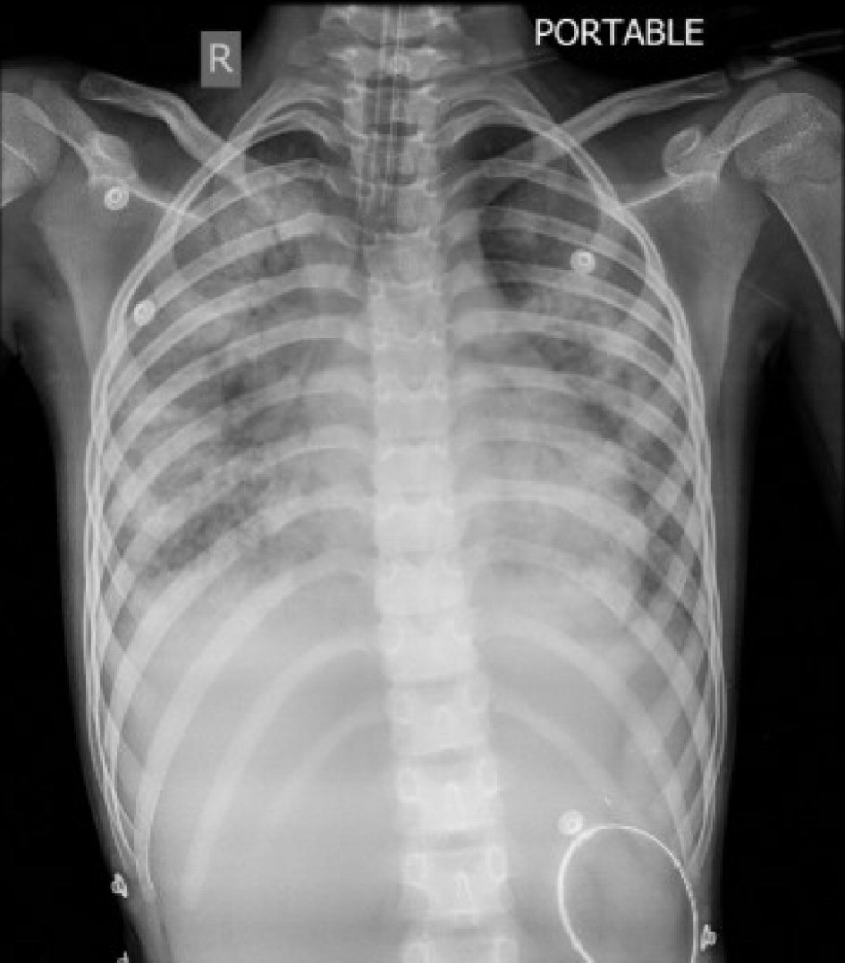 Click for large image | Figure 1. Chest X-ray at initial presentation. |
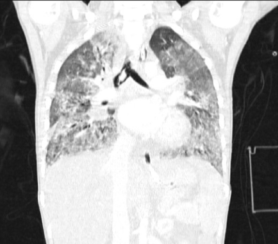 Click for large image | Figure 2. Computed tomography chest showing ground glass opacity and bilateral alveolar hemorrhages. |
She remained in the pediatric intensive care unit on ventilatory support. Five doses of pulse methylprednisolone (20 mg/kg/day) starting on day 4 of admission was given followed by prednisolone (2 mg/kg/day given for 7 days followed by 1 mg/kg/day for 7 days and then continued on 0.6 mg/kg/day) and hydroxychloroquine (4 mg/kg/day for 2 months). Her enteral feeds were initiated with gluten-free diet. Her clinical course was complicated by a urinary tract infection (urine culture was positive for extended-spectrum beta-lactamase (ESBL) Escherichia coli (E. coli)) and steroid-induced hypertension which were managed accordingly. She was managed with meropenem for a total of 14 days.
She improved dramatically with the treatment and was extubated 3 days after initiation of pulse methylprednisolone. There was gradual but complete resolution of her symptoms. She was discharged asymptomatic after 2 months of hospital stay. A follow-up after 1 month revealed her hemoglobin of 11.8 g/dL on low dose steroid (0.6 mg/kg/day) and gluten-free diet.
| Discussion | ▴Top |
LHS is an association of IPH and CD with very few case reports published in literature. IPH is an uncommon cause of diffuse alveolar hemorrhage with unknown pathogenesis. The response to immunosuppressive therapy indicates that an immune process may be involved. CD is an autoimmune enteropathy affecting individuals with genetic predisposition with ingestion of gluten [6]. The association or the causal relationship between both conditions is not clearly understood [6, 7]. There are a few hypotheses regarding the association of IPH and CD [8].While one states deposition of immune complexes on the basal membrane of alveolar capillaries, another considers it a cross-reaction of anti-reticulin antibodies with the alveolar basal membrane antigens, and a third hypothesis links adenovirus involving lungs along with gut.
The association of IPH and CD was first described by Lane and Hamilton in 1971 [9]. While this association is very rare, it is of clinical importance as a gluten-free diet for treatment of CD can lead to remission of symptoms of IPH. Complete resolution of symptoms has been reported in a series of cases [6, 10-15]. IPH is a rare cause of pulmonary hemorrhage in children. Data on pulmonary hemorrhage in children are insufficient as it is difficult to carry out any prospective study due to its rarity. In a recent study from India, IPH was found in 36.4% cases of pulmonary hemorrhage in children in a cohort of follow-up patients [12]. Reading et al [13] first reported improvement of pulmonary symptoms by gluten-free diet in IPH. In a comprehensive review literature by Singhal et al [10], in which 35 cases reported, 62.9% were of pediatric age group (< 18 years). It was also observed that pulmonary symptoms responding to gluten-free diet were higher in children (63.6%). In another study from India [16], the association of IPH and CD was found in 2.1% patients in non-diarrheal CD in the pediatric population. The emphasis for screening for CD is made in all patients with IPH even without gastrointestinal symptoms [17, 18].
The clinical course of IPH is variable. Diffuse alveolar hemorrhage is recurrent and the symptom severity can vary from blood-tinged sputum to severe and life-threatening hemorrhage requiring respiratory support. Continuous alveolar hemorrhage can lead to alveolar capillary block and can increase mortality if underlying cause is not treated. The authors did a systematic PubMed search of pediatric cases with a combination of IPH and CD. A total of 30 pediatric patients were reported on 24 occasions (Table 1 [6-9, 11, 13, 15, 17-33]). The mean age of presentation of all reported pediatric cases is 9.5 years, with standard deviation (SD) of 4.4 years. The most common presenting symptom was recurrent hemoptysis (70%) followed by anemia (63%) and cough (23%). Gastrointestinal symptoms were present in 20% of the reported cases. Dilated cardiomyopathy was seen in three cases [21, 24, 25], and retinitis pigmentosa in two cases [19, 33] reported as LHS. To the best of our knowledge based on literature available, our case appears to be the first reported pediatric case of LHS presenting with an acute episode of hemoptysis and fulminant respiratory failure with no previous constitutional symptoms. Gluten-free diet was advised in all the 30 reported cases. It is noteworthy that 42% of the pediatric cases showed improvement in pulmonary and other presenting symptoms with only gluten-free diet. Favourable outcome in pulmonary symptoms with gluten-free diet warrants early recognition of LHS.
 Click to view | Table 1. List of Pediatric Patients Reported in the Literature With a Combination of Idiopathic Pulmonary Hemosiderosis and Celiac Disease |
Conclusions
In conclusion, hemoptysis in children is a rare presenting complaint, and IPH is a diagnosis of exclusion. A high index of suspicion of IPH in a fulminant case of hemoptysis can be lifesaving. Prompt evaluation for CD in IPH is necessary as an association exists. Combined treatment with corticosteroids and gluten-free diet for LHS improves prognosis.
Acknowledgments
None to declare.
Financial Disclosure
None to declare.
Conflict of Interest
None to declare.
Informed Consent
Informed consent was obtained.
Author Contributions
AC, RH and RN have all made substantial contributions in the design of the work and drafting, final approval of the version and agreement to be accountable for all aspects of the work.
Data Availability
The authors declare that data supporting the findings of this study are available within the article.
| References | ▴Top |
- Pianosi P, al-sadoon H. Hemoptysis in children. Pediatr Rev. 1996;17(10):344-348.
doi pubmed - Tom LW, Weisman RA, Handler SD. Hemoptysis in children. Ann Otol Rhinol Laryngol. 1980;89(5 Pt 1):419-424.
doi pubmed - Fabian MC, Smitheringale A. Hemoptysis in children: the hospital for sick children experience. J Otolaryngol. 1996;25(1):44-45.
- Bidwell JL, Pachner RW. Hemoptysis: diagnosis and management. Am Fam Physician. 2005;72(7):1253-1260.
- Ioachimescu OC, Sieber S, Kotch A. Idiopathic pulmonary haemosiderosis revisited. Eur Respir J. 2004;24(1):162-170.
doi pubmed - Hammami S, Ghedira Besbes L, Hadded S, Chouchane S, Ben Meriem C, Gueddiche MN. Co-occurrence pulmonary haemosiderosis with coeliac disease in child. Respir Med. 2008;102(6):935-936.
doi pubmed - Khemiri M, Ouederni M, Khaldi F, Barsaoui S. Screening for celiac disease in idiopathic pulmonary hemosiderosis. Gastroenterol Clin Biol. 2008;32(8-9):745-748.
doi pubmed - Perelman S, Dupuy C, Bourrillon A. [The association of pulmonary hemosiderosis and celiac disease. Apropos of a new case in a child]. Ann Pediatr (Paris). 1992;39(3):185-188.
- Lane DJ, Hamilton WS. Idiopathic steatorrhoea and idiopathic pulmonary haemosiderosis. Br Med J. 1971;2(5753):89-90.
doi pubmed - Singhal KK, Janmeja AK, Sodhi R, Punia RS. Hemoptysis in patients of celiac disease with disproportionately severe anemia: tip of the iceberg? Multidiscip Respir Med. 2013;8(1):25.
doi pubmed - Sethi GR, Singhal KK. Pulmonary diseases and corticosteroids. Indian J Pediatr. 2008;75(10):1045-1056.
doi pubmed - de Silva C, Mukherjee A, Jat KR, Lodha R, Kabra SK. Pulmonary hemorrhage in children: etiology, clinical profile and outcome. Indian J Pediatr. 2019;86(1):7-11.
doi pubmed - Reading R, Watson JG, Platt JW, Bird AG. Pulmonary haemosiderosis and gluten. Arch Dis Child. 1987;62(5):513-515.
doi pubmed - Agarwal R, Aggarwal AN, Gupta D. Lane-Hamilton syndrome: simultaneous occurrence of coeliac disease and idiopathic pulmonary haemosiderosis. Intern Med J. 2007;37(1):65-67.
doi pubmed - Testa ME, Maffey A, Colom A, Aguero L, Roge I, Andrewartha MS, Teper A. [Pulmonary hemorrhage associated with celiac disease]. Arch Argent Pediatr. 2012;110(4):e72-76.
- Bhattacharya M, Kapoor S, Dubey AP. Celiac disease presentation in a tertiary referral centre in India: current scenario. Indian J Gastroenterol. 2013;32(2):98-102.
doi pubmed - Ertekin V, Selimoglu MA, Gursan N, Ozkan B. Idiopathic pulmonary hemosiderosis in children with celiac disease. Respir Med. 2006;100(3):568-569.
doi pubmed - Ostrom K, Syan R, Barber L, Miller D, Venkatasubramani N, Ravindra A, Jhaveri P, et al. Index of suspicion. Pediatr Rev. 2014;35(9):396-404.
doi pubmed - Keskin O, Keskin M, Guler E, Tutar E, Saygili O, Kucukosmanoglu E, Kor Y, et al. Unusual presentation: pulmonary hemosiderosis with celiac disease and retinitis pigmentosa in a child. Pediatr Pulmonol. 2011;46(8):820-823.
doi pubmed - Kamienska E, Urasinski T, Gawlikowska-Sroka A, Glura B, Pogorzelski A. Idiopathic pulmonary hemosiderosis in a 9-year-old girl. Eur J Med Res. 2009;14(Suppl 4):112-115.
doi pubmed - Yacoub M, Mahjoub H, Abroug S, Bousnina M, Harbi A, Essoussi AS. [Idiopathic pulmonary hemosiderosis, celiac disease and cardiomyopathy]. Arch Pediatr. 1994;1(6):587-590.
- Hartl D, Belohradsky BH, Griese M, Nicolai T, Krauss-Etschmann S, Roos D, Wintergerst U. Celiac disease and pulmonary hemosiderosis in a patient with chronic granulomatous disease. Pediatr Pulmonol. 2004;38(4):344-348.
doi pubmed - Nacaroglu HT, Sandal OS, Bag O, Erdem SB, Bekem Soylu O, Diniz G, Ozturk A, et al. Association of Celiac Disease With Idiopathic Pulmonary Hemosiderosis; Lane Hamilton Syndrome. Iran J Pediatr. 2015;25(5):e3312.
doi - Narula N, Rawal P, Kumar RM, Ram Thapa B. Association of celiac disease with cardiomyopathy and pulmonary hemosiderosis. J Trop Pediatr. 2010;56(3):201-203.
doi pubmed - Poddar B, Shava U, Srivastava A, Kapoor A. Severe heart failure, dilated cardiomyopathy and pulmonary haemosiderosis in coeliac disease: report of two cases. Paediatr Int Child Health. 2014;34(2):142-144.
doi pubmed - Paksu S, Paksu MS, Kalayci AG, Sancak R. Lane-Hamilton syndrome: association or coincidence. Indian Pediatr. 2012;49(3):243-244.
- Hendrickx GF, Somers K, Vandenplas Y. Lane-Hamilton syndrome: case report and review of the literature. Eur J Pediatr. 2011;170(12):1597-1602.
doi pubmed - Najada AS, Dahabreh MM. Pulmonary haemosiderosis in a 13-year-old girl with coeliac disease after 3 months on a gluten-free diet: case report and review of the literature. Ann Trop Paediatr. 2010;30(3):249-253.
doi pubmed - Pichardo C, Muinos W, Brathwaite C, Hernandez E. Pulmonary hemosiderosis associated with celiac disease: Lane Hamilton Syndrome. J Pediatr Gastroenterol Nutr. 2017;64(5):e133.
doi pubmed - Ploier R, Emhofer J, Dorninger L, Kranzl G, Feichtinger J, Muller KM, Brandtzaeg P. [Immunological aspects of a child with idiopathic pulmonary hemosiderosis and celiac disease]. Klin Padiatr. 1998;210(6):409-412.
doi pubmed - Hoca NT, Dayioglu D, Ogretensoy M. Pulmonary hemosiderosis in association with celiac disease. Lung. 2006;184(5):297-300.
doi pubmed - Panda PK, Sriranga R, Kaur K, Sood R. Lane Hamilton Syndrome. Indian J Pediatr. 2018;85(8):699.
doi pubmed - Villegas VM, Rachitskaya AV, Lam BL, McKeown CA, Berrocal AM. An unusual ophthalmic finding in Lane-Hamilton syndrome. J AAPOS. 2014;18(6):616-617.
doi pubmed
This article is distributed under the terms of the Creative Commons Attribution Non-Commercial 4.0 International License, which permits unrestricted non-commercial use, distribution, and reproduction in any medium, provided the original work is properly cited.
International Journal of Clinical Pediatrics is published by Elmer Press Inc.